-
Call Now
1800-102-2727

X rays - Production of X-rays, Properties of X-rays , Application of X-rays, practice problems, FAQs

Have your bones been fractured ever? When you go to the doctor, he will lay you down on a horizontal table of a big machine and click an image of your internal fracture. The machine on which you lay down is x- ray machine which produces the x- ray. When the x-rays pass from your body, the energy from X-rays is absorbed by various bodily components at various speeds. The X-rays penetrate through the body and are picked up by a detector on the opposite side, where they are processed into an image. Bones, which are dense and more difficult for X-rays to penetrate, appear as clear white regions on the image. Darker regions appear where there are softer tissues that are easier for X-rays to pass through, including your heart and lungs. This image is used to examine the crack and fracture of the bone. Let's learn more about the x-rays.

Table of content
- Introduction
- X-rays
- Production of X-rays
- Characteristics of X-rays
- Application of X-rays
- Practice problems
- FAQs
Introduction
Electromagnetic waves are defined as waves that can travel without a medium consisting of Electric and magnetic fields oscillating perpendicular to one another. Light waves, radio waves, infrared waves, X-rays, and other electromagnetic waves are a few examples of electromagnetic waves.
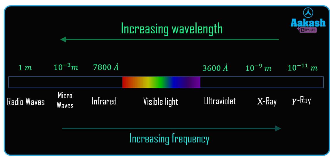
The electromagnetic spectrum is a range of frequencies, wavelengths, and photon energies for electromagnetic waves. The frequency ranges from 1 Hz to 1025 Hz, equivalent to wavelengths from a few hundred kilometres to a size smaller than an atomic nucleus. So in simple words, the electromagnetic spectrum can therefore be defined as the range of all types of electromagnetic radiation . In a vacuum, the speed of all electromagnetic waves is equal to the speed of light. However, different forms of electromagnetic waves will have different values for their respective wavelengths, frequencies, and photon energies. X-rays is the part of electromagnetic spectrum frequency ranging from the 1016 Hz to 1020 Hz.
X-rays
A category of extremely intense electromagnetic waves which is found between ultraviolet and gamma rays is known as X-rays. It is more energetic than UV rays but less energetic than gamma rays. It has a wavelength that varies from 0.01 to 10 nanometers, and frequency ranges between 1016 Hz to 1020 Hz.
German physicist Wilhelm Konrad Röntgen first saw X-rays in 1895 while studying the effects of electron beams, also known as cathode rays, in electrical discharges in low-pressure gases. In a stunning discovery, Röntgen demonstrated that a screen covered in fluorescent material and put outside of a discharge tube would glow even when insulated from the gaseous discharge's direct visible and ultraviolet light. He concluded that the screen fluorescence as a result of an invisible radiation from the tube passing through the air. This invisible radiation is called X- rays.
Production of X-rays
X-rays are often produced by three different processes: the acceleration of a charged particle, atomic transitions between distinct energy levels, and radioactive decay of particular atomic nuclei. Each method results in a specific X-ray radiation spectrum.
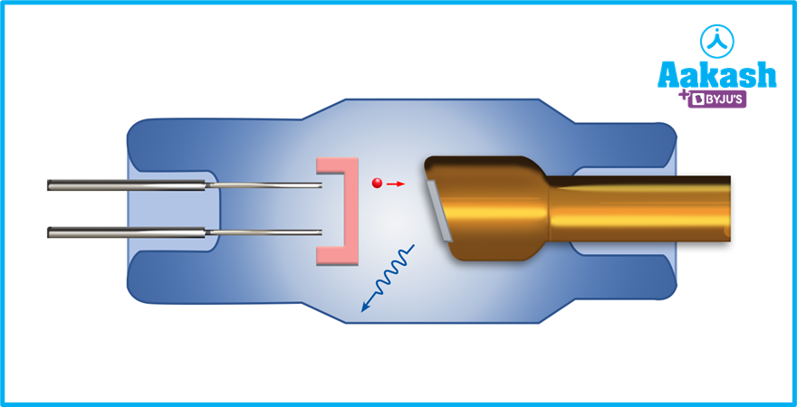
A beam of highly energetic electrons strikes a solid target in the X-ray tube, the most popular terrestrial source of X-rays. The rapidly travelling electrons in the beam are repeatedly deflected and slowed as they come into contact with the electrons and nuclei of the target atoms. A continuous spectrum of electromagnetic radiation with a peak intensity in the X-ray region, is produced by the beam electrons as a result of this rapid deceleration.
Characteristics of X-rays
The x-rays have the following properties:
- The wavelengths of the x-rays are quite small, ranging from 0.01 to 10 nanometers.
- X-rays travel at a similar speed of 300,000 km/sec as visible light.
- They can travel independently without the use of a media.
- X-Rays are produced with a high voltage.
- They are able to move through a vacuum.
Application of X-rays
The distinctive properties of X-rays, such as their capacity to pass through optically opaque materials, their atomic-scale wavelengths, and the high energy of individual X-ray photons, have led to a wide variety of commercial, medicinal, and scientific uses.
- Medical field
- X-ray radiography: Detects bone fractures, specific cancers and other abnormal masses, pneumonia, some types of traumas, calcifications, foreign objects, or dental issues.
- Mammography: A breast radiograph that is used to identify and diagnose cancer.
- Computed tomography (CT) : It is a conventional x-ray technology with computer processing to provide a series of cross-sectional images of the body that may later be combined to build a three-dimensional x-ray image. Doctors can observe body components from a variety of angles with CT images because they are more detailed than standard radiography.
- Fluoroscopy: x-rays and a fluorescent screen are used to provide real-time images of body motion or to observe diagnostic procedures, such as tracing the path of an injected or swallowed contrast agent.
- Therapeutic : By causing DNA damage to cancerous tumours and cells, X-rays and other high-energy radiation sources can be utilised to treat cancer.
- In Engineering field : It is possible to identify crystal structures in inorganic, organic, and biological materials using X-ray diffraction techniques also known as "X-ray crystallography".
- In Security : We have all used X-ray scanners to inspect our luggage in airports or metro stations. Without having to open the bag, X-rays disclose what is within and can identify metals, bombs, and other suspicious materials. Authorities can therefore be alerted to manual checks and take the necessary measures.
Practice problems
Q. How do X-rays in a typical X-ray machine get produced?
A. In X-ray tubes, X-rays are created when electrons that have been accelerated by a potential difference are directed at a target, usually a metal. The energy is transmitted to the metal as a result of this impact, and the metal's electron goes through a transition, as a result, releasing energy in the form of X-rays.
Q. What is spectroscopy?
A. The study of how light and other electromagnetic radiation are emitted and absorbed by materials based on the wavelength or frequency of the radiation is known as spectroscopy.
Q. Are the x- rays harmful for the body?
A. Yes, X-rays can injure the human body. This is why they must be handled carefully and with great caution when diagnosing medical conditions. Prolonged exposure to extremely powerful X-rays can harm the body's tissues and muscles.
Q. What are the x-rays?
A. X-rays are one of the types of the electromagnetic wave. The wavelength of the x-rays ranges from 0.01 to 10 nanometres and frequency ranging from 1016 Hz to 1020 Hz.
As like the other electromagnetic waves they can travel in the vacuum with speed of the light.
FAQs
Q. Can we see the x- rays?
A. X- rays can not be seen by naked eye. The only part of the electromagnetic spectrum which can be seen through the naked eye is the visible region.
Q. Which particle accelerates in X-ray emission tubes and releases X-rays as a result?
A. The electrons in X-ray emission tubes are accelerated by a very high voltage and then quickly slowed down by adding a metal plate. This process results in the conversion of electron kinetic energy into light energy and the emission of X-rays.
Q. Which of the following illnesses can be found with an X-ray?
- fever
- bladder infection
- pneumonia
- diarrhoea
A. A chest x-ray test can be used to identify pneumonia. The presence of white patches on the lungs indicates pneumonia in that person.
Q. What is the term used to describe the dental X-ray?
A. The word that is used to describe the dental X-ray is orthopantomography. X-rays are used in dentistry to identify illnesses, ingrowths, outgrowths, and abnormalities. It is employed to research the composition of teeth.