-
Call Now
1800-102-2727


Chromosomes: Structure, Types, Karyotype, Functions, Practice Problems and FAQs
What are factors which differentiates you from your siblings? It can be the colour of your eye, the colour and texture of your hair, the colour of your skin, the height or even the shape of your nose and ears. Right? Some of these features you might have inherited from your father and some from your mother. You might have even inherited features from your grandparents that are missing in your parents. The transferring of genetic characters from one generation to another is called heredity and is a really interesting field of study.
So how do we inherit these features? Heredity occurs due to transfer of genes that are present in chromosomes, from parents to offspring. The fundamental structural and operational unit of heredity is a gene. But the way genes are transferred from one generation to another does not always follow a similar pattern. That is why you are different from your siblings.
Genes are the special units of DNA and DNA are the building blocks of chromosomes. The chromosomes are present inside the nucleus of the cell. During the process of sexual reproduction, as the gametes from the parents fuse, the chromosomes carried by them form pairs with different combination of genes in the zygote which are responsible for all the genetic characters of the offspring. So every secret behind the character of an individual lies inside the chromosomes. By now you must be excited to know more about the chromosomes. Here we are going to discuss different types of chromosomes and let’s check out their functions.
Table of contents:
- Chromosomes
- Karyotype
- Special types of chromosomes
- Functions of chromosomes
- Practice Problems
- FAQs
Chromosomes
If we consider the structure of a nucleus, there will be protoplasm enclosed by a nuclear membrane which is also called Karyoplasm. It is a jelly like matrix. It contains water, lipids, proteins and dissolved ions and also has a nucleolus suspended in it along with chromatin.
Fig: Structure of nucleus

So what we are going to discuss now is about this chromatin which lies in the nucleoplasm.
Chromatin
It is also known by the names chromatin network, nuclear reticulum, or chromatin fibres. The nucleoplasm of a stained cell contains light-stained fibres that resemble threads which are referred to as chromatin. In a living, unstained cell, the chromatin network is not visible. The chromosomes are formed during cell division by the thickening of chromatin fibres. Higher organisms have a well-organised nucleus that contains a specific number of chromosomes with definite number and shape.
Fig: Condensation of chromatin to form chromosome

Chemical composition of chromatin fibres
- Continuous linear DNA duplex strand
- Associated basic proteins, histones or protamines
- Acidic or neutral histone proteins
- Small amount of RNA
- Some complex proteins, the enzymes DNA-polymerase and RNA polymerase
- Some phosphorus containing organic components
- Inorganic components such as salts
Types of chromatin fibres
There are two types of chromatin fibres - euchromatin and heterochromatin. This classification is based on the location and intensity of staining of chromatins in the nucleus of a cell undergoing interphase which is the longest preparatory phase of the cell cycle before cell division starts. Before entering mitosis, the cell grows during this time and duplicates its DNA.
Fig: Types of chromatin in the nucleus

Euchromatin
It is the metabolically or transcriptionally active form of chromatin. It is in the form of long chromatin fibres which are loosely packed. It is lightly stained. Under electron microscope the euchromatin has a bead on a string appearance. Each bead is called a nucleosome.
Fig: Beads on string arrangement of chromatin

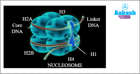
The major parts of a nucleosome are:
- Core particle
- Double stranded DNA fragment
- Linker DNA
- H1 protein
Core particle is an octamer composed of 8 histone molecules. These are two molecules each of H2A, H2B, H3, and H4.
Double stranded DNA is wrapped around the core particles. It is 146 nucleotide pairs long and forms 1.75 coils.
Linker DNA is about 60 base pairs long and it connects the two adjacent nucleosomes.
The H1 protein molecule is present in relation with each nucleosome. Along with the linker DNA, the H1 histone protein, joins the two nucleosomes.
Fig: Parts of nucleosome
Heterochromatin
They are the densely packed part of chromatin and they are darkly stained. It is of two types. They are constitutive heterochromatin and facultative heterochromatin.
Constitutive heterochromatin is present in all cells and all the stages of life cycle. It occurs near the centromeric region and telomeres of all the chromosomes.
Facultative heterochromatin is formed only in certain cells temporarily in certain stages of the life cycle. For example condensation of one of the two X chromosomes (XX) in female mammals to form Barr body represents heterochromatization. This condensed X chromosome is also called sex chromatin. Supercoiling or condensation of chromatin is called heteropycnosis.
Structure of chromosome
Now we know that the condensation of chromatin fibres will result in the formation of chromosomes. The major parts of a chromosome are the chromatids, centromere, kinetochore, telomeres, secondary constriction and matrix.
Chromatids
During prophase of mitosis, each chromosome duplicates and forms two sister chromatids that are connected by a centromere during cell division. The sister chromatids are identical to each other. The sister chromatids divide into individual chromosomes during anaphase.
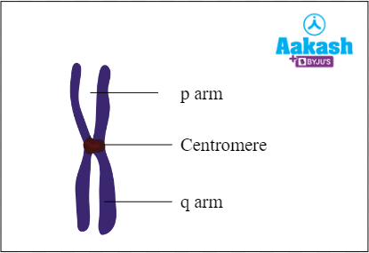
Centromere
The central portion of a chromosome is called centromere. The centromere links the two chromatids together. Centromere divides the chromosome into short p arm and long q arm. It is also called primary constriction.
Kinetochore
Proteinaceous disc shaped structure on the sides of the centromere is called kinetochore. It helps in cell division. Its role is to allow chromosomal mobility during the anaphase stage of cell division.
Telomere
The terminal end of the chromatids are called telomeres.
Fig: Structure of chromosome

Secondary constriction
Apart from the primary constriction, some metaphase chromosomes may have one or more than one secondary constriction. It can be found in sites other than primary constriction, mostly at terminal ends. Secondary constrictions are narrow areas of two types - joints and NOR (Nucleolar Organiser Region). Joints are involved in breaking and fusion of chromosome segments. NOR, as the name suggests, is responsible for the formation of nucleolus. Secondary constrictions can serve as markers because they are consistently in the same locations.
Part of the chromosome separated by secondary constriction is called satellite. Chromosomes having satellites are also known as SAT chromosomes. Satellites help in distinguishing between organisms and SAT chromosomes in which the secondary constriction is involved in nucleolus formation are known as nucleolar SAT chromosomes..
Fig: Chromosomes with a satellite

Matrix
The membrane enclosing each chromosome is known as a pellicle. The jelly-like substance found inside the pellicle is called matrix. It is made of non-genetic materials such as RNA, proteins and lipids.
Classification of chromosomes
If we observe the chromosome, we can see the two arms present on either side of the kinetochore on the centromere. The shorter arm is called the p arm and the longer arm is called the q arm. When the site of the primary constriction or centromere is clearly apparent during metaphase and anaphase, the chromosomal shape is typically visible. Chromosomes can be classified into four types based on the position of the centromere. They are:
- Metacentric
- Submetacentric
- Acrocentric
- Telocentric
Fig: Parts of chromosome

Metacentric chromosomes
These chromosomes have the centromere at the centre with two equal arms. Here the p arm = q arm.
Fig: Metacentric Chromosome
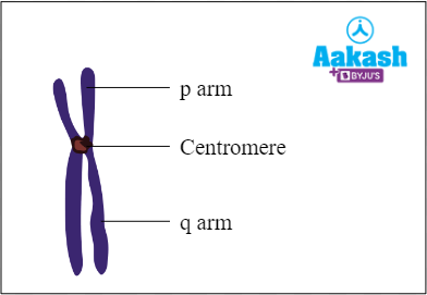
Sub metacentric chromosomes
These chromosomes have the centromere near the centre or we can say that they have a subterminal centromere with one shorter arm and one longer arm. Here the p arm < q arm.
Fig: Submetacentric Chromosome
Acrocentric chromosomes
These chromosomes have the centromere close to their ends forming one extremely short and one very long arm. Here the p arm < < q arm.
Fig: Acrocentric Chromosome

Telocentric chromosomes
These chromosomes have terminal centromeres. Here there is no p arm, only q arm is present.
Fig: Telocentric chromosome

Karyotype
A set of chromosomes with a set of fixed traits defines the members of a species of plant or animal. Number of chromosomes, relative size, centromere position, arm length, secondary constriction, and satellites are a few examples of traits of chromosomes in an organism. Karyotype represents the complete set of chromosomes (at metaphase stage), in any cell of a species or an individual organism, that are arranged according to length, centromere location and other traits. The diagrammatic representation of a karyotype is known as an idiogram. Karyotyping is the process of preparing a karyotype with the help of photographs of chromosomes.
Human karyotype
Human karyotype is a representation of the chromosomes present in a human cell. A human cell has 46 chromosomes of different shapes and sizes. Out of the four types of chromosomes that we have discussed so far, three can be observed in our human karyotype. Telocentric chromosomes are absent in humans. Then what about the rest of the types? Let’s check it out.
Chromosomes 1, 3, 16, 19 and 20 and X chromosome are metacentric. Chromosomes 2, 4 and 12 are sub-metacentric. Chromosomes 13, 14, 15, 21 and 22 and Y chromosome are acrocentric.
Fig: Human karyotype

Importance of karyotyping
After discussing the karyotypes and idiograms, there will be a definite question arising in your minds. What is the importance of karyotyping? Right? So let’s see what is the purpose of knowing the karyotypes of different organisms.
Karyotyping helps us:
- to find out the primitive or advanced features of an organism.
- to find out evolutionary relationships among different species.
- to find out any variation in individual chromosomes such as duplication and deletion through flow cytometry. The flow cytometry technique allows detection of difference as small as 1.5-4.0 Mbp (mega base pair).
Special types of chromosomes
Certain chromosomes in eukaryotic organisms are only present in specific types of tissues and are not found in other tissues. These chromosomes are referred to as giant chromosomes because of their size. They can be found in the suspensors of the embryo of several plants. The polytene chromosome and the lamp brush chromosome are the two different forms of giant chromosomes which are considered as the special type of chromosomes.
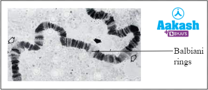
Polytene chromosome
C.G. Balbiani discovered polytene chromosomes in the salivary glands of Drosophila in 1881. Polytene chromosomes are found in the cells of larvae of Drosophila, mosquitoes and midges and are large chromosomes consisting of thousands of DNA strands.
Large chromosomes in polytenes are a result of their high DNA content. They are created through multiple rounds of chromosomal DNA replication during interphase without nuclear division. The resulting daughter chromatids do not split apart and remain joined together instead. The larval cells die during metamorphosis because they are unable to go through mitosis.
The interbands, which are a series of black bands that alternate with transparent zones down the length of the chromosome, are what distinguish polytene chromosomes from other types. The Balbiani ring is a large puff on the polytene chromosome. Another name for it is chromosomal puff. This chromosome is referred to as a salivary gland chromosome since it is found in the salivary gland.
Fig: Polytene chromosome
Lamp brush chromosome
Flemming discovered lamp brush chromosomes in 1882. It had a brush-like appearance. They are the largest known chromosome. We can observe the lamp brush chromosomes in both the giant nucleus of the unicellular alga Acetabularia and the oocytes of many vertebrates (except mammals) and invertebrates during the extended diplotene stage of the meiotic prophase. The chromosomal axis is formed by the highly condensed chromosome. These chromosomes serve as templates for intense RNA production which causes the extension of lateral loops of DNA.
Fig: Lamp brush chromosome

Functions of chromosome
- Sutton and Boveri were the first to propose that chromosomes play a function in heredity in 1902.
- The most significant role of chromosomes is to carry the DNA, which is the basic genetic material. DNA contains the genetic code for many biological processes. These processes are necessary for the development, maintenance, and procreation of the organisms.
- Chromosomes serve the additional purpose of shielding DNA from damage during cell division. Chromosomes are protected by proteins such as histones which shield them from both physical and chemical forces like enzymes.
- Spindle fibres connected to the centromeres contract during cell division and help in separating the sister chromatids and pulling them to the opposite poles of the cell. The exact distribution of DNA to the daughter nuclei is ensured by the splitting of the chromosomal centromeres and the contraction of the spindle fibres attached to them.
- Histone and non-histone proteins are found in chromosomes. These proteins control the action of genes.
Practice Problems
1. Which one of the following lacks DNA?
- Mitochondria
- Chloroplast
- Nucleus
- Lysosome
Answer: The nucleus is the organelle in a eukaryotic cell that stores the DNA that makes up the organism. The double-membraned organelle known as the nucleus is also referred to as the "brain of the cell." It manages every aspect of the cell's operations. Chloroplasts and mitochondria are double-membraned organelles that each have their own DNA. Circular double-stranded DNA can be found in both the mitochondria and chloroplasts. Lysosomes are single membraned sac-like organelles. They don't have any DNA. These are abundant in hydrolytic enzymes, which can break down a variety of dietary components, including proteins, lipids, and carbohydrates. As they can ingest damaged or dead organelles as well as the entire cell, they are often known as the suicide bags of the cell.
Hence the correct option is d.
2. Find the appropriate match between the chromosomal type and centromere position.
|
Chromosomes |
Position of centromere |
|
(A) Metacentric |
(1) At the tip |
|
(B) Telocentric |
(2) Almost near the tip |
|
(C) Acrocentric |
(3) At the middle |
|
(D) Submetacentric |
(4) Slightly away from the middle |
- (A)-(1), (B)-(2), (C)-(3), (D)-(4)
- (A)-(2), (B)-(4), (C)-(3), (D)-(1)
- (A)-(3), (B)-(1), (C)-(2), (D)-(4)
- (A)-(3), (B)-(2), (C)-(1), (D)-(4)
Answer: A chromosome is seen to include two identical halves during cell division, known as chromatids, which are held together at a location known as the centromere. Chromosomes can be divided into four groups based on where the centromere is located. Chromosomes that are metacentric are the chromosomes whose centromere is located in the middle (median). Chromosomes that are submetacentric have centromeres that are submedian, or located slightly away from the centre or the middle of the chromosome. Chromosomes which have their centromeres very close to one of the ends are known as acrocentric chromosomes (almost near the tip). The centromere of the telocentric chromosome is located at the chromosome's terminal end (at the tip).
Hence the correct option is c.
3. Spindle fibres attach to the
- cell membrane of the cell
- telomere of the chromosome
- ribosomes of the cell
- kinetochore of the chromosome
Answer: Protein-rich fibres called spindle fibres help in cell division. During mitotic and meiotic divisions, they are in control of sister chromatid segregation and chromosomal mobility. Sister chromatids are drawn toward their opposing poles by the action of spindle fibres, which cling to the kinetochore.
Hence the correct option is d.
4. What is the difference between the chromatin and chromosome?
Answer:
|
Chromatin |
Chromosomes |
|
Observed in interphase nucleus |
Observed during M phase of nuclear division |
|
Uncondensed state of nucleoprotein complex |
Condensed state of nucleoprotein complex |
|
Network of fine threads called chromatin reticulum |
A short, thick, cord like structure |
|
Controls the metabolism and other activities of the cell |
Transfer the genetic information to the daughter cells |
FAQs
1. What are chromonema and chromomere?
Answer: Chromonema is a coiled filamentous structure that resembles a thread and is where chromomeres are organised. The chromonema regulates chromosomal size and serves as a location for gene expression. The bead-like structures found on threads or chromonema are called chromomeres. Along the length of chromonema, these are arranged up in a row.
2. What is a FISH?
Answer: The Fluorescence In Situ Hybridization is known as FISH. It is the technique used to make the karyotypes of animals and plants. In FISH, the positions of specific DNA sequences or chromosomes can be located by using the probes that are labelled with fluorescent dye. The fluorescently labelled probe locates its matching sequence among the collection of chromosomes and binds to it. The chromosome and subchromosomal sites where the fluorescent probe binds can be observed with the aid of a specialised microscope.
3. What is the role of colchicine in karyotyping?
Answer: In order to obtain separated chromosomes during karyotyping, a mitotic inhibitor is used which can stop cell division. A typical mitotic inhibitor used in cytogenetic procedures like karyotyping is colchicine. Inhibiting the development of spindles and stopping the cell division in metaphase are the effects of colchicine.
4. What is a philadelphia chromosome?
Answer: The Philadelphia chromosome was the first frequently occurring genetic change discovered to be connected to a particular type of human cancer, which is the Chronic Myeloid Leukaemia (CML). It was initially discovered to be an abnormally small chromosome in CML cells. Later, a chromosomal rearrangement involving the BCR gene on chromosome 22 and the tyrosine kinase ABL gene on chromosome 9 was discovered to be the cause. Cells are hypothesised to develop uncontrollably as a result of the BCR-ABL fusion gene. The Philadelphia chromosome can be located in a range of ways, which can be utilised to diagnose CML and track a patient's clinical progress.