-
Call Now
1800-102-2727


Bacteria: Characteristics, Structure and Types of Bacteria, Heterotrophic Bacteria and its Classification, Practice Problems and FAQs
Do you know what a bacteria is? The first thing that comes to our mind when we hear of a bacteria is ‘diseases’. But did you know that not all bacteria cause diseases? In fact, there are many bacteria that live in our bodies and are responsible for our good health. Curd is an integral part of our daily diet because of its high nutritional value and health benefits. Did you know that it is produced when bacteria grows in milk? When lactic acid bacteria such as Lactobacillus feed on milk sugar lactose and produce lactic acid, they cause the milk protein casein to precipitate due to acidic pH and this precipitated protein forms the curd.
But this is just an example of a single bacterial species. Did you know that there are around 30,000 formally named species of bacteria and there could be many more which have not yet been identified? In fact, you will be amazed to know that these are one of the most primitive organisms to have evolved on earth and yet they have established themselves in every nook and corner of our planet and are pretty much omnipresent.
In this page, we will discuss all about bacteria, their structure, types, how they are classified and their significance.

GIF: Bacteria
Table of contents
- Kingdom Monera
- Bacteria
- Characteristics of bacteria
- Structure of bacteria
- Types of bacteria
- Heterotrophic bacteria
- Classification of heterotrophic bacteria
- Significance of bacteria
- Practice Problems
- FAQs
Kingdom Monera
All prokaryotes belong to the kingdom Monera. They are known as the oldest microorganisms on the earth which lack a well-defined nucleus and their genetic material lies naked in the cytoplasm. They lack any organelles that are membrane-bound. Kingdom Monera consists of unicellular organisms which are commonly found in a moist environment.
Classification of Monera
Kingdom Monera is classified into two categories:
- Archaea
- Eubacteria
Archaea
These are the ancient bacteria that can be found in extreme conditions, such as the marshes (methanogens), hot springs (thermoacidophiles), and salty places (acidophiles). They are also known as archaebacteria. These bacteria lack peptidoglycans in their cell wall. Instead of this, the cell wall is composed of pseudopeptidoglycans, glycoproteins, pure proteins, and polysaccharides. Their cell membrane is also formed of a monolayer of lipids that have long, branched hydrocarbon chains linked to glycerol. These structures enable archaebacteria to withstand extreme temperatures and pH.
Eubacteria
They are also known as true bacteria because the cell wall is rigid and is made up of peptidoglycan. These include cyanobacteria (photosynthetic blue-green algae) and heterotrophic bacteria, Mycoplasma, Rickettsia and Actinomycetes.
Bacteria
Eubacteria or bacteria are single-celled prokaryotic organisms which are highly specialised and occur everywhere around us. They are found in soil, in air, in water, as parasites in other organisms, hot springs, deep oceans, snow, etc. Some of these bacteria, such as the nitrogen fixing soil bacteria, are very useful while some can be extremely harmful pathogens. The reason why bacteria are universally distributed are -
- Extremely simple structure
- Small size and consequently large surface area to volume ratio. Small size is maintained by frequent cell division.
- Highly resistant endospore formation.
- Diverse modes of nutrition.

Fig: Habitat of Bacteria
Characteristics of bacteria
The bacteria possess the following salient features:
- They are unicellular and prokaryotic cells.
- They are found in a variety of shapes, sizes, and arrangements.
- The bacterial cell does not have a nucleus and membrane-bound organelles.
- The bacterial DNA is present naked in the cytoplasm in the form of a nucleoid and it is not packed in the form of chromatin as in eukaryotes.
- They have 70S ribosomes scattered in the cytoplasm.
- Respiratory enzymes are located on the inner surface of the cell membrane.
- Generally, the bacterial cell is 10 times smaller than the human cell.
- The diameter of a bacterial cell is approximately 1 µm.
- The outer covering of the bacterial cell is rigid and provides structural integrity.
- Peptidoglycans or murein make up the cell wall of bacteria.
- They have various modes of nutrition including photoautotrophic, chemoautotrophic or heterotrophic.
- Photoautotrophic forms have bacteriochlorophyll and chlorobium chlorophyll instead of a typical chlorophyll.
- The different shapes of bacterial cells serve as the characteristic features of bacterial species.
- Some of the bacterial cells contain external appendages, such as cilia and flagella.
- The food reserve in bacterial cells is in the form of glycogen and fat globules.
Structure of bacteria
The structure of bacteria is very simple because it is a single-celled microorganism that lacks a nucleus and other membrane-bound cellular organelles. Therefore, they are known as prokaryotic organisms. The bacterial cells have an outermost complex cell envelope which is composed of an outermost glycocalyx, then a rigid cell wall and an innermost cell membrane. If the glycocalyx is in the form of a thick and rigid mucilaginous sheath then it is known as a capsule. When the glycocalyx is in the form of a loose sheath it is known as a slime layer.
Other than this, the bacterial cell is composed of :
- Cytoplasm
- Nucleoid
- Ribosomes
- Mesosomes
- Plasmids
- Flagella or Pili
Cell wall
The cell wall of a bacterium is rigid and protects the cell from the outer environment. It is made up of a mucopeptide named peptidoglycan (or murein). It is a polymer made up of alternating subunits of N-acetyl glucosamine (NAG) and N-acetyl muramic acid (NAM). A small 3-5 amino acid long peptide side chain attached to NAM cross links with the peptide chain of another strand.
The cell wall of gram-positive bacteria is composed of a thick layer of peptidoglycan complexed with teichoic acid. It prevents osmotic lysis of the cell protoplast and gives cells structure and rigidity. On the other hand, the cell wall of gram-negative bacteria is composed of a thin layer of peptidoglycan which is surrounded by a layer of lipoproteins and lipopolysaccharides. The peptidoglycan layer prevents osmotic lysis and ensures the rigidity and shape of the cell.
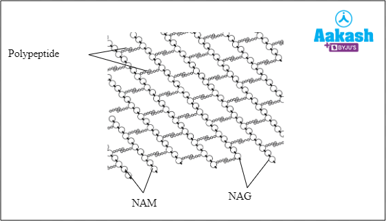
Fig: Peptidoglycan structure
Plasma membrane
The plasma membrane of bacterial cells is composed of phospholipids and proteins. It acts as a semipermeable barrier due to which it doesn’t allow all molecules to pass through. It is the site of energy production by cellular respiration as respiratory enzymes are located on it.
Nucleoid
The bacterial cell lacks a true nucleus and therefore, a nucleoid is present which is a region of cytoplasm where chromosomal DNA is located. It comprises one molecule of circular DNA duplex, which is supercoiled and stabilised with the help of RNA and non-histone proteins. It lies freely in the cytoplasm and is not bound by a nuclear membrane.
Plasmid
These are extrachromosomal hereditary determinants which can replicate independently. The plasmids may contain important genes such as fertility factors, antibiotic resistance genes, nif genes for nitrogen fixation, etc.
Ribosomes
These are described as the protein factories where protein synthesis takes place through a process called translation. They translate the genetic code from the mRNA to amino acids which are the building blocks of protein. The ribosome in bacterial cells is of the 70S type, made up of a smaller 30S subunit and a larger 50S subunit.
Mesosomes
Mesosomes are described as tubular-vesicular membrane structures that are formed by invaginations of the plasma membrane. These structures are more prominent in gram-positive bacteria as compared to gram-negative bacteria. These contain enzymes of the electron transport system and DNA replication. Mesosomes help in respiration, secretion and synthesis of cell wall material, receive DNA during conjugation and distribution of DNA to daughter cells during cell division.
Flagella
The bacterial flagella is an organ of locomotion which is composed of globular flagellin protein subunits arranged in a helical fashion. The diameter of each subunit is around 40Å-50Å.
Pili
These are hair like outgrowths present all over the surface of some Gram-negative bacteria. These are composed of helically arranged subunits of pilin protein and are meant for attachment. During conjugation, one or two special pili known as sex pili form a bridge between two bacteria for the transfer of DNA from the male (donor) bacteria to the female (recipient) bacteria.

Fig: Structure of bacteria
Classification of bacteria
Bacteria are further classified based on the following features:
- Shape
- Clusters
- Presence or absence of flagella and its distribution
- Reproduction
- Cell wall
- Nutrition
- Respiration
Types of bacteria based on the shape
On the basis of shape, bacteria are classified into five categories:
Coccus/Cocci |
Bacillus/Bacilli |
Spirillum/ Spirilla |
Vibrio |
Spirochete |
|
Spherical shaped Examples - Diplococcus (in pairs), Streptococcus (arranged in a chain), Staphylococcus (arranged in clusters) |
Rod-shaped Examples - E.coli, Bacillus anthracis |
Spiral shaped Examples- Spirillum voluntas, Treponema |
Comma shaped Examples - Vibrio cholerae |
Slender and spiral in shape Examples- Spirochaeta, Borrelia, and Leptospira |
|
Fig. Coccus/ Cocci |
Fig: Bacillus/ Bacilli |
Fig: Spirillum |
Fig: Vibrio/ comma shape |
Fig: Spirochete |


Types of bacteria based on clusters
On the basis of clusters, bacteria are classified into three categories:
-
Monococcus
These bacteria exist as a single spherical cell.
-
Diplococcus/Diplobacillus
In Diplococcus or Diplobacillus bacteria, the cells are arranged in a pair. Examples include Gram-negative bacteria, such as Neisseria spp, Moraxella catarrhalis and Gram positive bacteria, such as Enterococcus spp, etc.
-
Streptococcus/Streptobacillus
These are bacteria in which the spherical (coccus) or rod-shaped (bacilli) cells are arranged in a single plane in a chain like pattern. Examples of Streptococci are Streptococcus mutans, Streptococcus pyogenes, Streptococcus bovis, Streptococcus agalactiae, etc.

GIF: Types of bacteria based on clusters
Types of bacteria based on flagellation
On the basis of the absence or presence of flagella, bacteria are classified into the following categories:
Atrichous |
Flagella is absent in these types of bacteria Fig: Atrichous bacterium |
Monotrichous |
A single flagellum occurs at one end of the bacterial cell. Fig: Monotrichous bacterium |
Amphitrichous |
Single flagellum is present on both the ends of bacterium Fig: Amphitrichous bacterium |
Lophotrichous |
Acluster of flagella occur at one end of the bacterium. Fig: Lophotrichous bacterium |
Peritrichous |
The entire bacterial cell is covered in flagella. Fig: Peritrichous bacterium |
Amphilophotrichous |
A cluster of flagella present at each end of the bacterial cell. Fig: Amphilophotrichous bacteria |


Reproduction in bacteria
On the basis of reproduction, bacteria are classified into two types:
- Asexually reproducing bacteria
- Parasexually reproducing bacteria
Asexually reproducing bacteria
Asexual method of reproduction in bacteria occurs in two ways, one binary fission and the other spore formation.
The most typical means of bacterial reproduction is binary fission. Firstly, the chromosomal DNA replicates and then the bacterial cell divides into two daughter cells due to the formation of a transverse septum in the centre of each cell.
Fig: Binary fission in bacteria

Spores are highly resistant structures that can withstand high temperature, radiation, antibiotics, and chemicals. These are formed to evade unfavourable conditions. Spores may be formed within the cell (endospore) or outside the cell (exospore). Under favourable conditions, spores germinate into bacterial cells. This process is known as spore formation.
Fig: Types of spores in bacteria

Parasexually reproducing bacteria
Bacteria cannot reproduce sexually because there is no union of male and female gametes to create a diploid zygote. Genetic recombination involves the transfer of some genes from a bacterium to another bacterium. There are three methods of genetic recombination in bacteria, one is transformation, second is conjugation, and third transduction.
Transformation is the process through which bacteria acquire foreign DNA from their environment and change into a different kind. It was first discovered in Diplococcus by Griffith in 1928.
Fig: Transformation in bacteria

On the other hand, conjugation is the process in which F plasmid is transferred from one bacterium to another with the help of sex pilus (conjugation tube). It was discovered for the first time in E.coli by Beadle and Tatum.
GIF: Conjugation in bacteria

Transduction is a type of parasexual reproduction in which DNA fragments are transferred from one bacterium to another through bacteriophages.
Fig: Transduction in bacteria

Types of bacteria based on cell wall
On the basis of the cell wall, bacteria are classified into two categories:
- Gram-negative bacteria
- Gram-positive bacteria
Gram positive bacteria |
Gram negative bacteria |
|
Lipids, though in smaller amounts, are present in the single-layered cell wall which contains a thick layer of peptidoglycan. |
Cell wall has two layers -
|
|
After the Gram staining procedure, cells look blue or purple under a microscope. |
After the Gram staining procedure, cells look red under a microscope. |
|
Fig: Cell wall structure of gram-positive bacteria |
Fig: Cell wall structure of gram-negative bacteria |


Types of bacteria based on nutrition
Bacteria show an extremely high degree of metabolic diversity and can be autotrophic or heterotrophic in nature.
Autotrophs
These bacteria synthesise their own food. They can be of two types as follows:
|
Photosynthetic autotrophs |
Chemosynthetic autotrophs |
|
These bacteria synthesise food using light energy with the help of photosynthesis. They have specialised spherical bodies called chromatophores which contain the photosynthetic pigment bacteriochlorophyll. Photosynthesis carried out by these bacteria is non-oxygenic, that is, they do not produce oxygen. |
These bacteria oxidise chemical compounds to obtain chemical energy which is used for the synthesis of food. |
|
Examples include the following:
Fig: Photoautotrophic - Chlorobium |
Examples include nitrifying bacteria such as Nitrosomonas, Nitrococcus, etc, which oxidise nitrogenous compounds and obtain energy. Fig: Chemoautotrophic - Thiobacillus |


Heterotrophic bacteria
The heterotrophic bacteria cannot manufacture their own food. They receive their food from dead organic matter or living organisms.
|
Saprotrophic bacteria |
Parasitic bacteria |
Symbiotic bacteria |
|
The food source for these bacteria is decomposing and dead organic matter. |
They feed off of their living hosts while residing on or inside of them. |
Some nitrogen-fixing bacteria live in symbiotic association with the roots of some plants and derive nutrition from the plant. |
|
Example: E.coli Fig: Saprotrophic - E.coli |
Example: Clostridium tetani Fig: Parasitic - Clostridium tetani |
Example: Rhizobium Fig: Symbiotic - Rhizobium |


Types of bacteria based on respiration
On the basis of respiration, bacteria are classified into two types:
- Aerobic bacteria: Bacteria that respire in the presence of oxygen.
- Anaerobic bacteria: Bacteria that respire in the absence of oxygen.
Aerobic bacteria
Aerobic bacteria are further classified into two types:
|
Obligate aerobic |
Facultative aerobic |
|
These are strictly aerobic and die in the absence of oxygen. |
These are normally anaerobic bacteria but have the capability of aerobic respiration. |
|
Example: Bacillus subtilis |
Example: Most photosynthetic bacteria |
Anaerobic bacteria
Anaerobic bacteria are further classified into two types:
|
Obligate anaerobic bacteria |
Facultative anaerobic |
|
These are strictly anaerobic bacteria which die in the presence of oxygen. |
These are normally aerobic bacteria but can survive in the absence of oxygen. |
|
Example: Clostridium botulinum |
Example: Clostridium tetani |
Significance of bacteria
The significance of bacteria in our daily lives is unparalleled. While some bacteria can be extremely harmful pathogens, some can be extremely useful for us and our environment. Some significances of bacteria can be summarised as:
- The bacteria present in the digestive tract or gut are beneficial for health. This is because they help in the digestion of food.
- Some bacteria are capable of producing antibiotics and can be used for industrial production of antibiotics, for example, Streptomyces griseus produces the antibiotic streptomycin.
- They are used in the manufacturing of yoghurt and fermented food items. Lactobacillus is used in the production of curd.
- Saprotrophic bacteria play an important role as decomposers as they feed on dead and decaying organic matter and break down the complex molecules in such matter into simple inorganic molecules which are released into the environment. This helps in cycling of nutrients.
- Symbiotic bacteria such as Rhizobium help in directly converting atmospheric nitrogen into ammonia and thus helps in biological nitrogen fixation.
- Ammonifying and nitrifying bacteria in soil help in the conversion of nitrogen present in the atmosphere to ammonia, nitrites and nitrates which is available for the absorption by plant roots.
Practice Problems
- Which of the following categories of bacteria obtains nutrients from a living host?
- Photoautotrophs
- Saprotrophs
- Chemoautotrophs
- Parasitic bacteria
Solution: Autotrophic bacteria are those which prepare their own food by using either light energy (photoautotrophs) or chemical energy obtained by oxidising inorganic compounds (chemoautotrophs). Saprotrophic bacteria are heterotrophs which obtain readymade food from dead and decaying organic matter. Parasitic bacteria are heterotrophs which either live within or on the body of another living host and derive their nutrition from the host. Hence, the correct option is d.
2. Identify the organisms that have peptidoglycans and amino acids in their cell wall.
- Archaebacteria and eukaryotes
- Bacteria and cyanobacteria
- Eukaryotes and protists
- Monerans and protists
Solution: The cell wall of bacteria and cyanobacteria is composed of peptidoglycans and amino acids, such as muramic acid. Hence, the correct option is b.
3. Which of the following correctly describes the cell wall composition of Gram negative bacteria?
- Double layered cell wall with thick peptidoglycan layer
- Double layered cell wall with thin peptidoglycan layer surrounded by lipoproteins and lipopolysaccharides.
- Single layered cell wall with thin peptidoglycan layer
- Single layered cell wall with thick peptidoglycan layer and no lipids
Solution: The cell wall of Gram negative bacteria is double layered and has a thin layer of peptidoglycan surrounded by a thick layer of lipopolysachharides and lipoproteins. These cells appear red under the microscope after being stained using the Gram statining technique. Thus, the correct option is b.
4. Identify the type of bacterium in which a cluster of flagella is present on one side.
- Lophotrichous
- Amphitrichous
- Monotrichous
- Atrichous
Solution: In lophotrichous bacteria, a bunch of flagella is present on one side of the body. In amphitrichous, a single flagellum is present on both ends of the bacterium. In monotrichous, only one flagellum is present on one end of the bacterium, and in atrichous, the flagellum is absent. Hence, the correct option is a.
FAQs
- What are probiotics?
Answer: Good or beneficial bacteria are known as probiotics. They are living microorganisms with the intent of enhancing health when ingested or administered to the body. Yoghurt and other fermented foods, dietary supplements, and cosmetics contain them.
- Who was the first person to coin the term bacterium?
Answer: Bacteria were first seen by Anton van Leeuwenhoek in 1683 and he named them as animalcules. Ehrenberg was the first person to use the term ‘bacterium’.
- Name the largest bacterium.
Answer: The largest bacterium known is Thiomargarita magnifica, also known as a giant bacterium. The size of this bacterium is about 0.4 inches.
- Why do some bacteria have a rod shape?
Answer: The rod shape of a bacterium provides an inherent symmetry to them due to which they can store proteins at specific locations.
Youtube Video
Related Topics
|
Kingdom Monera: Archaebacteria, Cyanobacteria, Mycoplasma, Rickettsia, Actinomycetes, Practice Problems and FAQs |
|
Overview of Kingdom Monera, Practice problems, FAQs |
|
Kingdom Monera : Eubacteria - Habitat, Shape, Structure, Motility, Pili, Practice Problems and FAQs |
|
Kingdom Monera: Eubacteria - Nutrition, Respiration, Reproduction |