-
Call Now
1800-102-2727


Human digestive system, Practice Problems and FAQs
Everyone has their own favourite food. Right? Some like vegetarian food, others like non vegetarian food. But we have to take food to obtain energy to carry out all our life processes and daily activities. Any food we eat should be digested properly, then only our body can absorb it. You all know that overeating or eating quickly will cause indigestion of food. Then how is this caused? It is caused by various factors. One common factor is acid reflux. It is the backward flow of acid from your stomach into the oesophagus which irritates your throat. This can result in indigestion. To avoid this we must eat healthy food properly on time.

Fig: Healthy food
The process of digestion starts from the mouth. You know the way we chew our food can affect digestion. This is the reason why our parents used to tell us to chew our food properly and to eat slowly. All the parts of our digestive system play a role in the process of digestion of food. So what all parts are included in the human digestive system and how is the food digested?
From mouth to anus the food passes through many parts of the gastrointestinal tract (GIT) and all through this path many enzymes are also helping us to digest the food. So let’s take a deep dive into the details of the human digestive system in this article.
Table of contents
- Human digestive system
- Alimentary canal (GIT)
- Associated digestive glands
- Practice Problems
- FAQs
Human Digestive system
The human digestive system is made up of organs that collaborate to turn food into energy that the body can use and the elimination of indigestible remains. The human digestive system and nutrition are concerned with the food intake and energy expenditure of an organism. This crucial system enables living organisms to obtain energy from various sources. Before the nutrients in the food we eat are used to create energy, it is processed properly.
Parts of the human digestive system
Human digestive system can be divided into two main parts as follows:
- Alimentary canal or gastrointestinal tract (GIT)
- Associated digestive glands

Fig: Parts of the human digestive system
Alimentary canal
The gastrointestinal tract (GIT), also known as the alimentary canal, is a long, hollow, coiled, muscular, and glandular tube with a variable diameter. The alimentary canal measures between 6 and 9 metres in length. The digestive tract has two openings such as mouth (anterior one) and anus (posterior one). There are several specialised parts in the GIT. These regions carry out a variety of specialised tasks that aid in the digestion of food, absorption, its assimilation and elimination of undigested food.
Parts of the alimentary canal
The parts of the alimentary canal include the following:
- Mouth
- Oral or buccal cavity
- Pharynx
- Oesophagus
- Stomach
- Small intestine
- Large intestine
Mouth
The first specialised organ of the alimentary canal is the mouth. It is a transverse slit. The moveable top lip and lower lip provide protection to this opening. Both lips have mucous membrane lining on the inside and skin covering on the outside.

Fig: Mouth
Buccal cavity
It is located inside the mouth. It is a large area that is enclosed on three sides by the jaws, the throat below, and the palate above. The jaws hold the teeth while the throat supports the tongue. Buccal cavity is composed of three parts. They are as follows:
- Palate
- Tongue
- Teeth

Fig: Parts of buccal cavity
Pharynx
The space behind the soft palate is called the pharynx. It is a 12 to 14 cm long tube. It is considered as the common channel for food and air. It facilitates food swallowing. The muscles of the pharynx can contract. This increases and widens the pharynx's lumen, which facilitates swallowing. It is divided into three parts. They are as follows:
- Nasopharynx
- Oropharynx
- Laryngopharynx

Fig: Location of pharynx
Oesophagus
It is a muscular, thin tube-like structure. It extends from the posterior side of the neck and thorax, then pierces the diaphragm and enters the abdominal cavity. It is two cm wide and roughly 25 cm long. The oesophagus is also known as the food tube, food pipe, or gullet. It is divided into three parts. They are as follows:
- Cervical part - It is present in the neck region.
- Thoracic part - It is present in the thorax region.
- Abdominal part - It is present in the abdominal region.
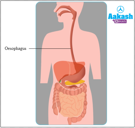
Fig: Location of oesophagus
A specialised movement known as peristalsis is used to move food from the throat to the stomach. Peristalsis is a series of wave-like muscle contractions that move food. The esophageal muscles block the passage of air into the digestive system. A band of muscles known as the cardiac sphincter surrounds the opening of the oesophagus into the stomach. It regulates the entry of food into the stomach.

GIF: Peristalsis
Stomach
The widest portion of the alimentary canal is the stomach. The location of the stomach is in the upper-left region of the abdomen and behind the diaphragm. It is a J-shaped organ. It is a hollow organ that holds food for up to four or five hours before breaking it down. Its upper side is more curved than its lower side. Due to its extremely convoluted interior surface, the stomach may fold up when empty and expand like a balloon when it is full of food. The greater omentum, a fold in the peritoneum attaches the stomach to the posterior abdominal wall.

Fig: Stomach
The stomach is divided into four parts. They are as follows:
- Cardiac
- Fundus
- Body or corpus
- Pylorus
Fig: Different parts of stomach

Small Intestine
The small intestine is a long, slender, and coiled structure that resembles a tube. In adults it is around 6.25 metres in length. It is divided into three parts as follows:
- Duodenum
- Jejunum
- Ileum
Fig: Small Intestine

Large Intestine
Considering its size, it is referred to as the large intestine. It is a nearly 2.5 m long tubular structure. The caecum, colon, and rectum are the three parts that make up the large intestine.
Fig: Parts of large intestine

Associated digestive glands
Digestive glands are the specialised structures that secrete digestive juices which help in the process of digestion. They include the following:
- Salivary glands
- Liver
- Pancreas
- Gastric glands
- Intestinal glands
Salivary glands
Saliva is released into the buccal cavity by salivary glands. There are three pairs of salivary glands as follows:
- Parotid glands
- Submandibular glands or submaxillary glands
- Sublingual glands
Fig: Salivary glands

Saliva
It is a watery substance produced in the mouth. A food related sight, scent, or thought can make more salivation. Saliva production ranges from 1 to 1.5 L per day. The pH of saliva is 6.8. It is formed of 98 - 99% of water. So there are four major components in the saliva. They are as follows:
- Water - It prevents drying of the mouth.
- Electrolytes - It acts as buffering agents.
- Salivary amylase - It is an enzyme that helps in the digestion of starch into maltose. It is also called ptyalin.
- Lysozyme - It is a naturally occurring enzyme that has antimicrobial properties.
Liver
It is considered as the largest gland in the human body. It weighs about 1.2 - 1.5 kg in adults. It is an important gland for digestion which is present in the upper right side of the abdominal cavity. It has a right and a left lobe. A ligament called the falciform ligament divides these lobes. Cells of the liver are known as hepatocytes. Bile is the digestive juice secreted by the liver.
Fig: Liver

Bile
Hepatocytes are the cells of the liver which secrete bile. It is stored in the gallbladder. The gallbladder is a pear-shaped organ that is attached by connective tissues to the posterior surface of the liver. It serves as a concentrating reservoir for bile. Bile consists of bile salts and bile pigments. Bile salts include sodium taurocholate, sodium glycolate, potassium taurocholate and potassium glycocholate. Bile pigments include bilirubin and biliverdin.
Fig: Gallbladder

Pancreas
It is the second largest gland in the human body. It is considered as a heterocrine gland. Hence along with hormones, the pancreas also secretes enzymes. The pancreas is divided into four parts such as the head, neck, body, and tail. The head can be seen in the duodenum's curvature.
Fig: External structure of pancreas

If we observe the internal part of the pancreas, we can see the grape-like clusters called acinar cells. This forms the exocrine part of the pancreas. An alkaline pancreatic juice that is secreted by the acinar cells aids in digestion. The pH of pancreatic juice is 8.4 - 8.8. Every day, 200 – 800 mL of pancreatic juice is secreted.
Water, sodium bicarbonates, trypsinogen, chymotrypsinogen, procarboxypeptidase, pancreatic lipase, pancreatic alpha-amylase, elastase, DNase, and RNase are the main components of pancreatic juice.
Fig: Internal structure of pancreas

Ducts of the liver and pancreas
The hepatopancreatic duct carries bile and pancreatic juice to the duodenum of the small intestine. Right and left hepatic duct originates from the respective liver lobes joins to form a common hepatic duct. It joins with the cystic duct from the gallbladder to form the bile duct. A number of small ducts within the pancreas join to form the main pancreatic duct or duct of Wirsung. The bile duct joins with the main pancreatic duct to form the hepatopancreatic ampulla (ampulla of Vater). It opens into the duodenum. This opening is guarded by the sphincter of Oddi.
Fig: Ducts of the liver and pancreas

Gastric glands
The epithelium of the stomach forms the tubular glands known as gastric glands. These glands release the digestive juice called gastric juice.
Fig: Gastric glands

Cells present in the gastric glands
There are several cells that make up the gastric glands. They are as follows:
- Chief cells or peptic cells - Secrete pepsinogen, pro rennin and gastric lipase.
- Oxyntic cells or parietal cells - Secrete HCl and intrinsic factor.
- Mucus cells or goblet cells - Secrete mucus.
- Enteroendocrine cells - Secrete serotonin and gastrin.
Intestinal glands
Epithelial cells undergo modification to become glands in the small intestine. It secrete the intestinal juice or succus entericus. This juice contains enzymes like lactase, amylase, maltase, isomaltase, nucleotidases, intestinal lipases, enterokinase, alpha dextrinase, nucleosidase etc. The small intestine has two different types of glands. They are as follows:
- Brunner’s gland - Secrete mainly mucus.
- Crypts of Lieberkuhn - Secrete digestive enzymes and mucus.
Fig: Intestinal glands

Significance of digestive system
The digestive system helps in the proper digestion, absorption and assimilation of food materials. It also helps in the elimination of undigested food materials. The proper functioning of this system is required for growth and development.
YOUTUBE VIDEO:
Practice Problems
Q1. In a restaurant, two friends are having dinner. One of them starts choking and coughing out of nowhere. This results from the improper movement of ______________.
A. diaphragm
B. neck
C. tongue
D. epiglottis
Solution: The respiratory and digestive systems share a channel called the pharynx. It allows the food and air to pass through it. The pharynx normally allows air to enter the larynx and trachea. The food or beverage must enter the oesophagus rather than the larynx when consumed. This is made possible with the help of the epiglottis, a cartilaginous flap that covers the opening of trachea while swallowing. The epiglottis can occasionally fail to cover the opening of the trachea, the glottis when a person is eating or drinking too quickly or talking while eating. This will lead the food or drink to enter into the trachea. When this happens, the person chokes and coughs to clear the food or liquid from their lungs. Hence the correct option is d.
Q2. Which of the following best describes how food should travel through a human digestive system?
A. Mouth → Pharynx → Buccal cavity → Oesophagus → Small intestine → Stomach → Large intestine → Anus
B. Mouth → Buccal cavity → Pharynx → Oesophagus → Stomach → Small intestine→ Large intestine → Anus
C. Mouth → Buccal cavity → Oesophagus → Pharynx → Stomach → Large intestine → Small intestine → Anus
D. Mouth → Buccal cavity → Stomach → Pharynx → Oesophagus → Small intestine → Large intestine → Anus
Solution: Mouth, Buccal Cavity, Pharynx, Oesophagus, Stomach, Small intestine, Large intestine, and Anus, is the correct order for food to go through the alimentary canal. The first section of the alimentary canal is the mouth. Food is consumed through the mouth. The palate, tongue, and teeth are all parts of the buccal cavity. Here, the food has been broken and combined with saliva. The pharynx serves as a common food and air channel. A muscular structure called the oesophagus transports food from the pharynx to the stomach. The upper left portion of the abdomen contains a muscular pouch called the stomach. It breaks down proteins into smaller molecules while churning the food. The longest section of the alimentary canal is the small intestine. Here, food digestion is completed. This is also the region where food absorption happens. In the large intestine, the undigested food is compressed into faeces, which are eventually expelled through the anus. Hence the correct option is b.
Q3. Which of the following is not included as a part of the large intestine?
A. Colon
B. Rectum
C. Ileum
D. Caecum
Solution: The large intestine is a portion of the GIT. The caecum, colon, and rectum are the three parts that make up the large intestine. It is 1.5 metres long. The caecum is a 6 cm long, pouch-like structure. The ascending colon, transverse colon, descending colon, and sigmoid colon are the four areas of the colon. The rectum is where the sigmoid colon opens. Rectum is the last 18 - 20 cm of large intestine and it opens into the 4.2 cm long anal canal. Anus refers to the opening of the anal canal. The highly coiled area of the small intestine is called the ileum. Hence the correct option is c.
Q4. Match the following cells present in the gastric glands with their secretion.
|
A. Peptic cells |
i) Mucus |
|
B. Parietal cells |
ii) Pepsinogen |
|
C. Goblet cells |
iii) Hydrochloric acid |
|
D. Enteroendocrine cells |
iv) Serotonin |
A. A - i, B - ii, C - iii, D - iv
B. A - ii, B - iii, C - i, D - iv
C. A - i, B - iii, C - iv, D - ii
D. A - ii, B - iii, C - iv, D - i
Solution: The epithelium of the stomach forms the tubular glands known as gastric glands. These glands release the digestive juice called gastric juice. There are several cells that make up the gastric glands. They are the chief cells or peptic cells, oxyntic cells or parietal cells, mucus cells or goblet cells and enteroendocrine cells. Chief cells or peptic cells secrete pepsinogen, pro rennin and gastric lipase. Oxyntic cells or parietal cells secrete HCl and intrinsic factors. Mucus cells or goblet cells secrete mucus. Enteroendocrine cells secrete serotonin and gastrin mainly. Hence the correct option is b.
FAQs
Q1. What are transverse rugae?
Answer: The buccal cavity's roof is divided into two sections. The term soft palate refers to the area of the roof that is posterior while hard palate refers to the area that is anterior. The hard palate has transverse folds known as rugae. Bones support them, so that they can hold the food that is being masticated. The soft palate assists in swallowing and has a smooth exterior.
Q2. Describe the Waldeyer's ring?
Answer: The Waldeyer's ring, which is located in the upper digestive tract, is a cluster of lymphoid tissue or follicles. Tonsils, adenoids, and other lymphoid tissue are components of the Waldeyer's ring. It has lymphocytes, which are immunological cells that support the defences against infection and illness. Four tonsillar structures are present. They are the pharyngeal, tubal, palatine, and lingual tonsils.
Q3. How quick is the digestive process in humans?
Answer: It takes between six and eight hours for the food to move through the stomach and small intestine after we eat. The food is digested here. In the large intestine water is absorbed and then the undigested food is ultimately eliminated from here. For food to pass completely through the colon, it takes roughly 36 hours.
Q4. Are you able to survive without a stomach?
Answer: We can survive without a number of different organs. But the fact that you can also survive without a stomach. The primary job of the stomach is to store and digest food, so that it may be passed on to the intestines. But with a little assistance, the body can adapt to bypass this process. This indicates that the duodenum serves as a direct route from the oesophagus to the intestine. This makes it possible for the pancreatic and bile ducts to continue draining into the duodenum and digesting food so that nutrients can still be absorbed.
Q5. Which food requires the most time to digest?
Answer: Bacon, steak, lamb, whole milk, hard cheese, and nuts take the longest time to digest. Our body needs four hours on an average to digest these food items. Even while we sleep, the digestive process continues.
Related Topics
|
Parts of digestive system: Anatomy and Histology, Practice Problems and FAQs |
|
Digestive glands: Salivary glands, Sjögren's Syndrome, Liver, Pancreas, Gastric glands, Intestinal glands, Practice Problems and FAQs |
|
Absorption and assimilation of digested products: Absorption in the small intestine, buccal cavity, stomach and large intestine, Assimilation, Egestion, Calorific value, Practice Problems and FAQs |
|
Buccal cavity: Structure and Role in digestion, Deglutition, Practice Problems and FAQ’s |
|
Stomach: Structure, Layers of Stomach, Role in Digestion, Practice Problems and FAQs |
|
Small intestine: Structure and Role in digestion, Absorption in small intestine, Practice Problems and FAQs |
|
Large intestine: Structure and contribution in digestion, Practice Problems and FAQs |