-
Call Now
1800-102-2727

Nucleus, Practice Problems and FAQs

We all have some favourite possessions which we consider precious. This can either be due to their material worth or the emotional attachment we share with those possessions. It is our natural instinct to preserve such possessions and keep them locked away for them to be safe. Right? So we look for the safest place to hide the treasures which have a huge significance in our lives.
Did you know, initially our Earth was a RNA world? RNA world hypothesis states that earth began with a single RNA molecule which had the ability to replicate and make copies of itself. Then later it evolved into an advanced and stable molecule which can carry the genetic information generation after generation; that was DNA.
In the biological world, DNA is the most precious thing which is needed for the perfect functioning of the living organism. It is responsible for the synthesis of proteins which are the building blocks of life. So protecting the DNA is essential. During the early years of evolution, the primitive single-celled organisms which existed did not have special means to protect their DNA and were hence more vulnerable. But as evolution took its course, a more evolved structure developed which ensured better protection to the DNA. As a result, a special organelle called nucleus was formed by the fusion of vesicles around individual chromosomes to form a membrane. These membranes then fused with each other and formed a complete single nucleus.
GIF: Synthesis of protein
Hence we can compare the nucleus and the DNA inside with the oyster protecting the precious pearl inside it. In this article we are going to discuss
Table of contents:
- Nucleus
- Structure of a nucleus
- Function of a nucleus
- Practice Problems
- FAQs
Nucleus
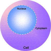
The most essential double membranous organelle which directs and controls all the activities of a cell is called the nucleus. Nucleus is known as the brain of the cell as it controls the whole cell and its functions. It is capable of doing so due to the presence of the genetic materia, i.e, DNA within it. It is a part of the endomembrane system, which includes other organelles like endoplasmic reticulum, Golgi apparatus and lysosomes.

Fig: Nucleus inside the cell; microscopic view
History of discovery
The nucleus was first observed by the father of optical microscopy, Antony van Leeuwenhoeck. He observed nuclei in red blood cells of amphibians in 1710. Then 1781 Felice Fontana observed nuclei in eel skin cells. In 1802, Franz Bauer pointed out the nucleus in his sketch of an orchid.
But it was Robert Brown, in 1831, who observed a structure that was uniformly present in the centre of all the plant cells he observed. He called it ‘Nucleus’.

Fig: Robert Brown
Types of cells on the basis of nucleus
The nucleus of all organisms are not similar to each other. There are different types of cells based on the presence or absence of a nucleus and also on the basis of the number of nucleus present inside it.
Classification of cells on the basis of presence or absence of nucleus
On the basis of presence or absence of well developed nucleus, there are two types of cells as follows:
- Prokaryotic cell
- Eukaryotic cell
On the basis of this, biologists classified the organisms into prokaryotes and eukaryotes.
Prokaryotic cell
In organisms like bacteria, archaebacteria and blue-green algae, the nucleus with a distinct nuclear membrane is absent. They have their genetic material in a region inside the cell called nucleoid which lies naked in the cytoplasm. Such cells are called prokaryotic cells.

Fig: Prokaryotic cell
Eukaryotic cell
In all other higher living organisms, the nucleus is well organised in the cells. Such cells are called eukaryotic cells.

Fig: Eukaryotic cell
Classification of cells on the basis of number of nucleus
There are different types of eukaryotic cells on the basis of the number of nuclei present inside the cell. The cells can be as follows:
- Enucleate
- Mononucleate
- Binucleate
- Multinucleate
Enucleate cells
The eukaryotic cells without a nucleus are called enucleate cells. Those cells will have only a limited life span and they are also unable to divide. Eg: RBC of mammals, sieve tubes of vascular plants.

Fig: Enucleate cells
Mononucleated or monokaryotic cells
The cells with a single nucleus are called mononucleated or uninucleate or monokaryotic cells. Generally most of the cells are uninucleate.

Fig: Plant cell
Binucleate cells
The cells with two nuclei are called binucleate or dikaryotic cells. Paramecium is an example. In this organism, one nucleus is small and called a micronucleus, the other one is large and is called a macronucleus. Both the nuclei have different functions. Micronucleus is for the storage of hereditary information for the next generation. Macronucleus is the one which governs the metabolic activities of the cell.

Fig: Paramecium
Multinucleate
The cells with many nuclei are called multinucleate or coenocytic cells or syncytium. This type of cells can be seen in the fungal hyphae and in certain algae. In animals, multinucleate condition is seen in muscle fibres and epidermis of Ascaris.

Fig: Multinucleate cells
Shape of the nucleus
Generally the nucleus is round. But it can be cylindrical or elliptical. A horseshoe shaped or lobed nucleus can be found in monocytes. Trilobed nucleus is found in neutrophils. Both monocytes and neutrophils are types of white blood cells. Highly branched nuclei can be observed in the silk spinning cells of insect larvae.

Fig: Different types of nucleus
Size and position of the nucleus
Size of the nucleus varies in different cells of an organism and in different organisms of different species. It may vary from 5 to 25 μm. Usually the nucleus can be seen in the central portion of the cell, but in plant cells it is shifted to one end of the cell due to the presence of highly developed vacuole inside the cell.
Chemical composition of nucleus
The major components of the nucleus are as follows:
- DNA (9-12%)
- Basic proteins or histones (15%)
- RNA (5%)
- Acid proteins (65%)
- Mineral ions (Ca2+, K+, Na+)
Structure of a nucleus
Since the nucleus is the one of the organelle that carries the genetic material, during cell division the nucleus also gets divided to transfer the genetic material to the newly formed cell. So the structure of the nucleus will be different in different phases of cell division. Hence here we are going to discuss the structure of an interphase nucleus or the non dividing nucleus. It can be distinguished into the following parts:
- Nuclear envelope
- Nuclear matrix
- Nucleoplasm
- Nucleolus
- Chromatin reticulum
Let’s discuss more about the structure of a nucleus.

Fig: Structure of a nucleus
Nuclear envelope
A double membrane structure covering the nucleus and separating the nuclear material from cytoplasm is called a nuclear envelope or karyotheca. It was first demonstrated by O. Hertwig in 1893 as a nuclear membrane. The name nuclear envelope was given by Anderson. It is visible only during the interphase stage of the cell cycle. The major function of a nuclear envelope is the communication with the cytoplasm. A nuclear envelope is composed of the following parts:
- Nuclear pores
- Outer membrane
- Perinuclear space
- Inner membrane

Fig: Parts of nuclear envelope
Nuclear pores
The nuclear envelope is not continuous. The numerous pores present on the nuclear envelope are called nuclear pores. The diameter of a nuclear pore ranges from 600 to 1000 Å. It was described by Callan and Tomlin in 1950. A cylindrical and hollow structure called annulus is plugged inside the nuclear pore. It is covered with a fine diaphragm. Nuclear pores allow the exchange of selective materials through it. From cytoplasm, the ribosomal units come out to the cytoplasm through nuclear pores. Proteins synthesised in the cytoplasm enters the nucleus through these pores.

Fig: Structure of nuclear pore
Outer membrane
It is also known as ectokaryotheca. It is the membrane which is in contact with the cytoplasm of the cell. It is continuous with a rough endoplasmic reticulum, hence associated with ribosomes.
Perinuclear space
The outer and inner membranes of the nuclear pore are separated by fluid filled perinuclear space. It is continuous with the lumen of endoplasmic reticulum.
Inner membrane
It is also known as endokaryotheca. It is a smooth membrane and it is in contact with the nucleoplasm. The inner surface of the inner membrane bears fibrillar material.
The nuclear envelope has a thickness of 90 Å and it has a composition similar to the plasma membrane of the cell. It is formed of two lipo protein membranes on either side of the perinuclear space.

Fig: Structure of nuclear envelope
Nuclear matrix
The meshwork of intermediate filaments formed of protein called laminin is the nuclear matrix. It is also known as nuclear lamina. It is attached to the inner surface of the inner nuclear envelope. It provides mechanical support to the nucleus.

Fig: Nuclear lamina in the nucleus
Nucleoplasm
The transparent semifluid substance or protoplasm enclosed by the nuclear membrane is called nucleoplasm. It is also called nuclear sap, karyoplasm or karyolymph. It is a jelly-like matrix, which contains water, lipids, proteins and dissolved ions. It also has nucleolus suspended in it along with chromatin. The major function is to provide rigidity to the nucleus and also support the chromatin material.

Fig: Nucleoplasm along with other parts of the nucleus
Nucleolus
The dense and spherical structure in nucleoplasm is called nucleolus. It is a thin thread-like network of nucleo-protein fibres. It is not bound by a membrane. It was first described by Fontana in 1781, later Wagner made a detailed study in 1840. The term nucleolus was used by Bowman in 1848.
It is the site of ribosome assembly. It is associated with the nuclear organising region or NOR of particular chromosomes which is represented by the secondary constriction in chromosomes. The number of nucleoli also varies depending on the type of cells. A diploid cell contains two or three nucleoli. The size of the nucleoli depends on the ribosomal requirements of a cell. The components of a nucleolus are as follows:
- DNA
- RNA
- Basic Histone proteins
- Non-histone proteins
The primary function of a nucleolus is the synthesis of rRNA and ribosomal assembly but it also plays an important role in the cell division.

Fig: Nucleolus
Chromatin reticulum
The stained cells contain lightly stained thread like fibres, and it is referred to as chromatin fibres, chromatin network, or nuclear reticulum. The chromatin network is not visible in a living, unstained cell.
Walther Flemming, in 1879, used basic stains to study the cell division in cells of Salamander. He observed some intensely stained parts in the nucleus and called them ‘Chromatin’.

Fig: Walther Flemming and his observations
The condensation of chromatin fibres forms the thick chromosomes and this happens during cell division. Higher organisms contain a specific number of chromosomes with definite number and shape, because they have a well-organised nucleus.

Fig: Condensation of chromatin to form chromosome
Types of chromatin
According to the level or intensity of staining, there are two forms of chromatin that exist:
- Euchromatin
- Heterochromatin
Euchromatin
The loosely packed and lightly stained chromatin is called euchromatin. It is the metabolically or transcriptionally active form of chromatin. Euchromatin has a bead on a string appearance, where each bead is called a nucleosome which is composed of a core of eight histone molecules wrapped around by a DNA strand. The major parts of a nucleosome are as follows:
- Core particle
- Double stranded DNA fragment
- Linker DNA
- H1 protein
Heterochromatin
The densely packed and darkly stained chromatin is called euchromatin. Due to its highly coiled structure, heterochromatin is not metabolically or transcriptionally active. The two types of heterochromatin are constitutive heterochromatin and facultative heterochromatin.
Constitutive heterochromatin is that heterochromatin which is present in all cells at all stages of the cell cycle. It usually occurs near the centromere, telomere and the ends of certain genes. It consists of repetitive nitrogenous bases.
Facultative heterochromatin is that heterochromatin which develops secondarily and is meant for inactivating chromatin or DNA regions that are nor required in certain cells or stages of life cycle.

Fig: Euchromatin and heterochromatin
Functions of nucleus
Since nucleus is the brain of cells, it performs many important functions which are as follows:
- The chromatin material is supported by the nucleoplasm of the nucleus.
- Nucleus can control the synthesis of enzymes required and thereby controls all the metabolic activities.
- The synthesis of DNA, RNA and ribosomal subunits takes place inside the nucleus.
- Nucleolus plays an important role in the cell division and synthesis of ribosomes.
- The inheritance of characters from parents to offspring is controlled by nucleus.
- Cell division is controlled by the nucleus.
Practice Problems
1. In which of the following pairs of organisms we can observe a nuclear material without a nuclear membrane?
- bacteria and green algae
- cyanobacteria and red algae
- bacteria and cyanobacteria
- mycoplasma and green algae
Solution: Prokaryotes have a nuclear material which is referred to as ‘naked’. This means that the nuclear material is not enveloped by a nuclear membrane. Bacteria, cyanobacteria (blue-green algae) and mycoplasma are prokaryotes. Alage is a eukaryotic organism. Genetic material is well enclosed by a distinct nuclear membrane in eukaryotes and they are said to have a true nucleus. Hence the correct option is c.
2. Select the correct statement from the following.
- The energy production machinery is present in nucleus
- All eukaryotic cells contain a nucleus at every stage of development
- On the outer nuclear membrane ribosomes can be observed
- Both b and c
Solution: A double membraned organelle containing the genetic material is called nucleus in a eukaryotic cell. Nucleus is the largest cell organelle in an animal cell. When observed in a non-dividing phase, the nucleus shows the nuclear membrane, chromatin, nucleoplasm and the nucleolus. The inner nuclear membrane does not have ribosomes present on it. But, the outer nuclear membrane has ribosomes on its surface. Perinuclear space is the space between the two membranes. The eukaryotic cells contain a well-defined nucleus. The mature RBCs do not have a nucleus at maturity. This is done to accommodate more space to transport gases. The mitochondria are also called the ‘powerhouse of the cell’ as these are the organelles where energy is synthesised in the form of ATP. It is the main site of aerobic respiration. The bonds in food substances are broken down in the presence of oxygen to release energy. The liberated energy is stored in the form of ATP. Hence the correct option is c.
3. Which of the following is true about the nuclear envelope?
- Consists of a single membrane with pores
- Consists of two membranes without pores
- Consists of two membranes without pores through and substances cannot pass through it
- Consists of two membranes with pores through and substances can pass through it
Solution: The nucleus is one of the cell organelles that carries the genetic material and during cell division the nucleus also gets divided to transfer the genetic material to the newly formed cell. So the structure of the nucleus will be different in different pages of cell division. The structure of an interphase nucleus or the non dividing nucleus can be distinguished into nuclear envelope, nuclear matrix, nucleoplasm, nucleolus and chromatin reticulum. A double membrane structure covering the nucleus and separating the nuclear material from cytoplasm is called a nuclear envelope or karyotheca. The major function of a nuclear envelope is the communication with the cytoplasm. A nuclear envelope is composed of nuclear pores, outer membrane, perinuclear space and inner membrane. The nuclear envelope is not continuous. The numerous pores present on the nuclear envelope are called nuclear pores which allow the exchange of selective materials through it. From cytoplasm, the ribosomal units come out to the cytoplasm through nuclear pores. Proteins synthesised in the cytoplasm enters the nucleus through these pores. Hence the correct option is d.
4. In which of the following cellular organelles the ribosomal RNA is produced?
- Golgi apparatus
- Mitochondria
- Cytoplasm
- nucleolus
Solution: The dense and spherical structure in nucleoplasm is called nucleolus. It is a thin thread-like network of nucleo-protein fibres. It is not bound by a membrane. It is the site of ribosome assembly. The non-membranous ribonucleoproteins (RNA and proteins) particles occurring in large numbers inside the cell are called ribosomes. The nucleolus is associated with the nuclear organising region or NOR of particular chromosomes. The primary function of a nucleolus is the synthesis of rRNA but it also plays an important role in the cell division. Hence the correct option is d.
FAQs
1. Do all cells have the same number of nucleoli?
Answer: A striking nuclear organelle which is present in all eukaryotes is nucleoli. The major function of nucleolus is to accelerate rRNA synthesis, processing, assembly of pre-ribosomal subunit etc. The number of nucleoli varies among different species. The number is fixed among the members of the same species. In a human diploid cell, we can observe only one nucleolus. But ten tiny nucleolus appear immediately after cell division. Then they coalesce into a single large nucleolus.
2. What are the differences between the nucleoplasm and cytoplasm?
Answer: Nucleoplasm and cytoplasm have differences in their position, structures present in it and the functions.
|
Features |
Nucleoplasm |
Cytoplasm |
|
Position |
Present inside the nucleus |
Present between the cell membrane and nucleus |
|
Structures present |
Nucleoli and chromatin fibres |
Cell organelles and cell inclusions |
|
Functions |
It is a part of the nuclear skeleton, helps in the formation of spindle fibres during cell division |
Glycolysis, protein synthesis, exchange of materials between the cell organelles |
3. How does RBC survive without a nucleus?
Answer: Immature erythrocyte or RBC is known as erythroblast. It contains a nucleus. When RBC matures they eject their nucleus and some more organelles to carry more haemoglobin and thus more oxygen. But they live around 100-120 days by using the proteins produced by the erythroblasts initially. The amount of oxygen required for the metabolism of RBC is very less, hence they can carry more oxygen to the tissues. Their biconcave shape also helps for this purpose.
4. What is the chemical composition of a chromatin?
Answer: The chromatin is composed of, continuous linear DNA duplex strand, associated basic proteins, histones and protamines, acid or neutral histone proteins, small amount of RNA, some complex proteins, the enzyme DNA polymerase and RNA polymerase, some phosphorus containing organic components and inorganic components such as salts.
YOUTUBE LINK: https://www.youtube.com/watch?v=eFul6cULbzU