-
Call Now
1800-102-2727

Tissue System: Ground Tissue System, Practice Problems and FAQs

Unity in diversity is the major component in every successful community. If different people with different skills and ideas come together to complete a job, they can not only finish it faster but also more efficiently. Inside a plant there is a unity in diversity among the tissues. Different types of tissues can together perform a particular function. For example the transportation in the plant body is done by both xylem and phloem in the vascular bundles. The mechanical strength of the plant is done by the collenchyma and sclerenchyma cells.
These tissues are present on the different parts of the plants. So on the basis of the structure and location of tissues present in plants are classified as the epidermal tissue system, ground tissue system and vascular or conducting tissue system.This classification was proposed by Sachs in 1975.
Do you know which one among these three tissue systems forms the bulk of the plants? It is the ground tissue system. Most of the tissues which we are familiar with are included in the ground tissue system and that is what we are going to discuss in this article.
Fig: Types of tissue systems

Table of contents:
- The ground tissue system
- Ground tissue system in dicot stems
- Ground tissue in monocot stems
- Ground tissue in dicot roots
- Ground tissue in monocot roots
- Ground tissue in leaves
- Practice problems
- FAQs
The ground tissue system
The ground tissue system is considered to include all the tissues except epidermis and vascular bundles. It is derived from the ground meristem. It forms the major part of the plant body. It is composed of simple tissues like parenchyma, collenchyma and sclerenchyma. The ground tissue system is a multilayered structure. The parenchymatous cells with intercellular spaces will be thin walled.
This tissue system is primarily responsible for providing support to the plant and may also help in storage of food. The ground tissue system in the roots stores food which is responsible for the formation of edible roots like carrots and turnips. Chlorenchyma cells in the ground tissues are responsible for the preparation of food, since chloroplast is present in them. The hypodermis protects the underlying layers and the endodermal cells, cortical cells and medullary rays contribute to conduction of water and nutrients.
The ground tissue system is mainly composed of hypodermis, cortical cells, endodermis, pericycle, conjunctive tissue and pith.
Fig: Ground tissue system of dicot stem

Hypodermis
Hypodermis lies under the epidermis of dicot stems. It may occasionally be regarded as the cortex's outermost layer. Sclerenchyma cells or collenchyma cells may make up the hypodermis. These cells can often be modified to provide additional structural support or to store food or water.
Cortex
Large, thin-walled parenchyma cells from the ground tissue system make up the majority of the cortex, which exhibits little to no structural differentiation. Cortical cells of young plants have chloroplast and can prepare food. Cells of the cortex also contain leucoplast for storage of starch grains. The cortex is responsible for transportation of water and salts from the root hairs to the centre of the root.
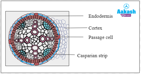
Endodermis
Endodermis is made up of tightly packed live cells. It has casparian strips (lignified thickenings) on both the tangential and radial walls. The casparian strip is water-resistant and helps the plant to regulate the amount of water and minerals it absorbs from the soil. Although an endodermis with casparian strip is always found in roots, it can also be found in the stems and leaves of some vascular plants.
Fig: Casparian strip

Some endodermal cells will have no casparian strips and those cells are called passage cells or transfusion cells. These cells allow radial diffusion of water and minerals through the endodermis.
Fig: Passage cells
Pericycle
The pericycle is a few layers thick and is formed of thick walled parenchymatous cells. The pericycle lies between the endodermis and the vascular tissues. The formation of lateral roots initiates from the pericycle. It also helps in secondary growth by forming the cork cambium.
Fig: Pericycle and formation of lateral roots from it

Conjunctive tissue
Parenchymatous cells which lie between the xylem and the phloem are called conjunctive tissues. If the tissue is parenchymatous, conjunctive cells serve as food storage units. After getting sclerified, they offer mechanical strength.
Fig: Conjunctive tissue

Pith
Pith is usually made of parenchyma cells and is located at the centre of the root. The pith is small and inconspicuous in dicot roots but it is large and conspicuous in monocot roots. The size of the pith varies in the anatomy of dicots and monocots.
Fig: Pith

Ground tissue system in dicot stems
A dicot stem consists of hypodermis, cortex, endodermis, pith and medullary rays. Hypodermis is the layer just below the epidermis. It is made up of collenchyma or sclerenchyma cells depending upon the plants. In plants, tissue of unspecialised cells lying between the hypodermis and the endodermis is cortex. Endodermis will usually be single layered rectangular cells lying just below the cortical layers. Endodermis are made up of starch grains, hence called starch sheath. A pericycle is a thin layer of plant tissue between the endodermis and the phloem. Pith is composed of undifferentiated parenchyma cells, which help in the storage of nutrients in young plants. Medullary rays are the strips of parenchyma which are present between vascular bundles of the dicot stem. They separate xylem and phloem bundles.
Fig: Ground tissues of dicot stem

Ground tissue in monocot stems
A monocot stem consists of hypodermis and ground parenchyma. It is not differentiated as cortex, endodermis, pericycle and pith. Vascular bundles are scattered in the ground tissues of monocot stems. The ground meristem is largely responsible for storage of food.
Fig: Ground tissues of monocot stem

Ground tissue in dicot roots
In the ground tissue system of a dicot root, many layers are present. They are cortex, endodermis, pericycle, pith, and conjunctive tissue. Casparian strips are present on the tangential and radial walls of endodermis. Passage cells are also present. Formation of vascular cambium is a major function of the pericycle of a dicot root.
Fig: Ground tissues of dicot root

Secondary growth occurs in dicot root by the formation of vascular cambium. The vascular cambium in dicot root arises from the conjunctive tissues present between the vascular bundles. The conjunctive tissues become meristematic and then they will form the cambial layers. Secondary meristems are formed in the roots after the tissues of the primary plant body have differentiated. The vascular cambium is responsible for increasing the diameter of roots resulting in the formation of woody tissues.
Fig: Formation of cambium from pericycle

Pith of a dicot root is small, inconspicuous and made up of parenchyma cells. Sclerenchymatous or parenchymatous conjunctive tissues are also present.
Ground tissue in monocot roots
The ground tissue system of a monocot root consists of different layers such as the cortex, endodermis, pericycle, and pith.
Fig: Ground tissues of monocot root

The cortex of monocot root lies inside the epiblema (outermost layer of the roots) and it is not well differentiated. Few outer layers become thick walled and suberised to form exodermis. Exodermis protects the older roots after decaying of epiblema. Young endodermal cells have casparian strips.They also possess passage cells. The pericycle produces lateral roots. A large pith is present in the centre. It consists of thin or thick walled parenchymatous cells having intercellular spaces.
Ground tissue in leaves
Leaves consist of mesophyll tissue in their ground tissue. These are thin-walled chloroplasts containing parenchymatous cells which lie between the upper and the lower epidermis of the leaf.
Mesophyll cells are differentiated into palisade parenchyma and spongy parenchyma in dicot leaves. But in monocot leaves, the ground tissue is not differentiated into palisade and spongy mesophyll.
Fig: Ground tissue in dicot leaf

Practice Problems
Q 1. Which of the following layers has a deposition of suberin in the anatomy of root or stem?
a. Epidermis
b. Endodermis
c. Pith
d. Pericycle
Answer: The endodermis layer of roots has a deposition of water-impermeable, waxy material called suberin. It is deposited on the radial and tangential walls of the endodermal cells and is called casparian strips.
Hence the correct option is b.
Q 2. Assertion: Ground tissue in monocot stems is made up of large, conspicuous and parenchymatous cells.
Reason: Storage of food is one of the functions of ground tissues of monocot stem.
Which of the following statements are correct according to the assertion and reason given above?
a. Both assertion and reason are true and the reason is a correct explanation of the assertion
b. Both assertion and reason are true but the reason is not a correct explanation of the assertion
c. The assertion is true but the reason is false
d. Both the assertion and reason are false
Answer: A monocot stem consists of hypodermis and ground parenchyma. It is not differentiated as cortex, endodermis, pericycle and pith. The cells comprising the ground meristem are large, conspicuous and parenchymatous in nature. This is because they are primarily involved with storage of food. Here the assertion is correct and reason is the correct explanation for the assertion.
Hence the correct option is a.
Q 3. What are the primary functions of ground tissues?
Answer: Ground tissue system is composed of simple tissues like parenchyma, collenchyma and sclerenchyma. This tissue system is primarily responsible for providing support and food storage. The ground tissue system in the roots also stores food which is responsible for the formation of edible roots like carrots and turnips. Chlorenchyma cells in the ground tissues are responsible for the preparation of food, since chloroplast is present in them. The hypodermis is responsible for protection of underlying layers and the passage cells, medullary rays and cortical cells also help in water conduction.
Q 4. What is the difference between the ground tissue system of dicot and monocot leaves?
Answer: The major difference in the ground tissues of dicot and monocot plants are observed in their leaf anatomy. Leaves consist of mesophyll tissue in their ground tissue. In dicot leaves mesophyll is differentiated into palisade parenchyma and spongy parenchyma. But in monocot leaves, the ground tissue is not differentiated into palisade and spongy mesophyll.
FAQs
Q 1. Does the cortex contain chlorophyll?
Answer: The cortex is a tissue of unspecialized cells that lies between the epidermis (surface cells) and the vascular, or conducting, tissues of stems and roots in plants. Chloroplasts may be present in a few of the outer cortical cells. As a result, the tissues of the outermost cortex shows the highest levels of photosynthetic activity and oxygen release rates.
Q 2. What are prosenchyma cells?
Answer: Prosenchyma cells are starch-containing parenchyma cells with lignin-lined cell walls, such as those seen in Bougainvillaea stems. The plant becomes more rigid due to prosenchyma. The term "transfer cell" also refers to prosenchyma.
Q 3. Who discovered casparian strips?
Answer: The Casparian strip was discovered in the mid-nineteenth century, and it contributed to our understanding of the Endodermis of plant roots. Robert Caspary, a German botanist, first identified the endodermis of a plant's root in 1865, discovering that its cell wall was thicker and naming it Schuchtzscheide. The thicker area of it was later called the Carls Belt, after Casbury, by researchers. The word "Caspary'schen fleck" first appeared in literature in the 1870s, and it was later referred to as the Casparian strip. Researchers separated the Casparian strip from the root of plants for the first time in 1922 to analyse its composition.
Q 4.What does the spongy mesophyll in a leaf do?
Answer: Spongy tissue is a component of the mesophyll in plants and lies below the palisade cells of the leaf, as seen in the transverse section of a leaf. The function of the spongy mesophyll is to allow for the exchange of gases such as CO2 which is required for photosynthesis.
YOUTUBE LINK: https://www.youtube.com/watch?v=GjfjE5B-P1w
Related Topics
|
Permanent tissues: Types of permanent tissues, Simple permanent tissues, Practice Problems and FAQs |
|
Epidermal tissue system: Epidermis, Stomata, Epidermal appendages, Practice problems and FAQs |
|
Plant vascular system, Types of vascular bundles, Practice problems and FAQs |