-
Call Now
1800-102-2727


NMR Spectroscopy – Definition, Principle, Instrumentation, Working, Detection, Practice Problems and FAQ
We often look at the photographs of our childhood and cherish them. Sometimes, our parents challenge us to identify our siblings or relatives from old and treasured photographs. We try to look for those striking features of resemblances. Imaging memories has been an age-old custom that treasures the golden past.
Identification of several molecules from our body, biological/physiological systems, drugs, and molecules belonging to our world at large has also taken the help of various imaging techniques. Scanning for a molecule’s structural features by identifying interaction patterns of its atomic nuclei when subjected to electromagnetic radiofrequency (RF) waves of a certain resonance frequency, is the generalised basis of NMR spectroscopy. An MRI scan basically uses this concept.

Doctors often prescribe patients take an MRI scan to diagnose abnormalities or tumours in certain parts of the body. This is an effective technique for measuring certain brain chemicals to distinguish between a brain abscess and a brain tumour. It is also used to identify fractures in the hip and pelvis as well as anomalies in the female reproductive system, and aid medical professionals in assessing sprains and anomalies of the joints such as knee cartilage or ligament tears etc.
Without further ado, let’s get back to learning and cracking down on NMR spectroscopy!
TABLE OF CONTENTS
- NMR Spectroscopy – Introduction
- NMR Spectroscopy – Principle
- NMR Spectroscopy – Characteristic Features
- NMR Spectroscopy – Chemical Shift
- NMR Spectroscopy – Instrumentation
- NMR Spectroscopy – Working and Detection
- NMR Spectroscopy – Shielding and Deshielding
- NMR Spectroscopy – Applications
- Practice Problems
- Frequently Asked Questions – FAQ
NMR Spectroscopy – Introduction
Nuclear magnetic resonance (NMR) spectroscopy allows scientists to analyse molecules by capturing the interaction of radiofrequency (RF) electromagnetic radiation with the nuclei of molecules put in a high magnetic field.
The end of 1945 saw the first experimental discovery of nuclear magnetic resonance (NMR), almost simultaneously with work groups led by Edward Purcell of Harvard University and Felix Bloch of Stanford University. The initial report on the first NMR spectra appeared in the same issue of Physical Review in January 1946. The 1952 Nobel Prize in Physics was shared by Bloch and Purcell for their work on nuclear magnetic resonance spectroscopy.
Zeeman originally noticed the peculiar behaviour of certain nuclei when exposed to a high magnetic field towards the end of the nineteenth century, but it wasn't until the 1950s when NMR spectrometers were made commercially accessible, that the so-called "Zeeman effect" was really put to use.
- NMR is a study method that makes use of some atomic nuclei's magnetic characteristics.
- It makes use of the nuclear magnetic resonance phenomena and offers thorough details regarding the kinetics, reaction state, and chemical environment of molecules.
- For organic chemists, nuclear magnetic resonance (NMR) spectroscopy is a vital analytical technique.
- It can provide information about the molecule's structure, physical and chemical characteristics of atoms or molecules as well as the sample's composition and purity.
- One of the NMR techniques that organic chemists utilise the most frequently is proton-NMR represented as Spectroscopy. Another example is. Basically, any sample of the molecule where nuclei show spin can be detected by NMR.
- It is possible to determine the structure of a molecule by observing how the protons in it respond to the surrounding chemical environment.
NMR Spectroscopy – Principle
Local magnetic fields surrounding atomic nuclei can be observed using the spectroscopic method known as NMR.
The sample is put in a magnetic field, and the nuclear magnetic resonance (NMR) signal is generated by radio waves excitation of the sample's nuclei, which is detected by sensitive radio receivers.
The intramolecular magnetic field of the atoms of a molecule can alter the resonance frequency, providing information about a molecule's electronic structure and its many functional groups.
In contemporary organic chemistry, NMR spectroscopy is the only reliable way to identify monomolecular organic molecules since the fields are distinctive or highly specific to particular compounds.
- The NMR principle states that all nuclei are electrically charged and that many nuclei have spin.
- When an external magnetic field is given, an energy transfer from the base energy to a higher energy level is possible.
- When an external magnetic field is provided, energy can be transferred from lower energy levels to higher energy levels.
- Energy is transferred at a wavelength that matches the radio frequency.
- Additionally, when the spin returns to its initial base level, energy is released at the same frequency. Therefore, the processing of the NMR spectrum for the concerned nucleus is produced by measuring the signal that fits this transfer.
Three successive processes typically make up the NMR principle.
- The polarisation (or alignment) of magnetic nuclear spins in a strong and constant magnetic field, B0, that is being applied.
- The alteration of this nuclear spin alignment caused by a weakly oscillating magnetic field, commonly known as a radio-frequency (RF) pulse.
- Detection and analysis of the electromagnetic waves emitted by the sample’s atomic nuclei that are generated due to this disruption caused at the sample's nucleus.
NMR Spectroscopy – Characteristic Features
- A spinning charge produces a magnetic field.
- Two spin states (up and down spin respectively), and , are present in the presence of an external magnetic field (B0).
- In contrast to the higher energy spin state, the magnetic moment of the lower energy state is in opposition to the external field.
- The intensity of the external magnetic field determines how little the energy difference between the two spin states is, which is always extremely little.
- For NMR spectroscopy, strong magnetic fields are essential. Powerful magnets with fields ranging from 1-20 T (Tesla) are used in modern NMR spectrometers. The energy difference between the two spin states is less than even at very high fields.
- This tiny energy differential (E) is often expressed for nmr purposes as a frequency in MHz (106 Hz), ranging from 20 - 900 Mz, depending on the intensity of the magnetic field and the particular nuclei under investigation.
- It is possible to excite a group of nuclei in the state to the higher spin state by irradiating a sample with radio frequency (RF) energy that precisely matches the spin state separation of that group of nuclei.
- Most importantly, this electromagnetic radiation belongs to the radio waves zone (radio frequency).
- NMR spectroscopy is thus the least energy-demanding probe available for studying molecular structure.
- At a certain magnetic field intensity, the energy gap between the two spin states for spin 12 nuclei will be proportional to their magnetic moments.
- The relation between magnetic moment frequency, magnetic moment, spin number (I) and Planck's constant is denoted as
NMR Spectroscopy – Chemical Shift
The chemical shift is the resonance frequency of an atomic nucleus in relation to a standard in nuclear magnetic resonance (NMR) spectroscopy. A molecule's structure may frequently be determined by looking at the position and volume of chemical changes.
.
- Chemical shift is defined as the difference between the signal from the reference molecule and the resonance frequency of the spinning protons present in the sample molecule’s atoms.
- One of the most significant characteristics that may be used to determine molecular structure is nuclear magnetic resonance chemical shift.
- NMR spectroscopy can also identify a variety of nuclei, including the proton (1-Hydrogen), Carbon-13, Nitrogen-15, and many more. The most popular are and . A magnetic field is created by nuclei that possess a spinning cloud of charge. Proton NMR is most commonly used.
- A magnetic field may produce various energy levels and resonance frequencies because some atomic nuclei have magnetic moments (also known as nuclear spin).
- The local geometry (binding partners, bond lengths, angles between bonds, etc.) and, consequently, the local magnetic field at each nucleus, often affect the electron distribution of the same kind of nucleus ( ). The spin energy levels reflect this (and resonance frequencies).
- Chemical shift refers to differences in electron distribution that cause changes in nuclear magnetic resonance frequencies of the same kind of nucleus. It is denoted by the symbol δ.
- The magnitude of the chemical shift is expressed in relation to a reference frequency or reference sample, often a molecule with a minimally perturbed electron distribution.
NMR Spectroscopy – Instrumentation
There are nine main components to an NMR spectrometer.
- Sample holder: Glass tubes measuring 8.5 cm long and 0.3 cm in diameter serves as the sample holder.
- Magnetic coils: When current travels through a magnetic coil, a magnetic field is produced.
- Permanent magnet: It aids in generating a uniform magnetic field between 60-100 MHz.
- Sweep generator: Adjusts the magnetic field's already-applied strength.
- Radiofrequency Transmitter: This device sends out a brief, strong radio wave pulse. It aids in the detection of receiver radio waves by radiofrequency.
- Radio frequency detector: It aids in locating unabsorbed radio frequencies.
- Recorder: It keeps track of the NMR signals that the RF detector picks up.
- Readout System: A computer that records the data is called a readout system.

NMR Spectroscopy – Working and Detection
- In a magnetic field, place the sample.
- Create NMR signals by using radio waves to excite the sample nuclei into nuclear magnetic resonance.
- With the use of sensitive radio receivers, these NMR signals are found.
- The intramolecular magnetic field surrounding an atom in a molecule alters its resonance frequency.
- This provides information on a molecule's electrical structure and specific functional groups.
- Monomolecular organic chemical identification may be done with certainty using nuclear magnetic resonance spectroscopy.
- This technique gives information on a molecule's reaction state, structure, chemical environment, and dynamics.
Resonant Frequency: It indicates the absorption energy and the signal intensity, which are inversely related to the magnetic field's strength. When exposed to a magnetic field, NMR active nuclei absorb electromagnetic radiation at a frequency specific to the isotope.
Spectral data collection: A nuclear magnetic resonance response is produced after the sample is excited by a radiofrequency pulse. Since it is so faint, only radio receivers with high sensitivity can pick it up.
NMR Spectroscopy – Shielding and Deshielding
NMR spectroscopy is the best method to determine the structure of molecules. Greater resistance to the applied magnetic field is produced by hydrogen atoms that have a higher electron density. The H-atom thus encounters a less magnetic field and is able to resonate at a lower frequency. For this H-atom, the peak on the NMR spectrum would move upfield. We refer to these H-atoms as being shielded.
The H-atom would experience a stronger magnetic field and would resonate at a higher radio frequency if it were surrounded by substances that lessen the electron cloud. This occurrence is known as de-shielding.
- Here, the chemical shift of the protons in CH4 and CH3Cl may be used as an example. Because chlorine is an electronegative element, it will attract electrons to it, deshielding the hydrogen nucleus. The chemical shift (in ppm) will thus be greater in the latter. Since the hydrogen nucleus is protected in the case of CH4, the peak is seen on the lower ppm side.
NMR Spectroscopy – Applications
NMR spectroscopy is important in the study of organic chemistry. Additionally, it is one of the strongest and most adaptable methods utilised in qualitative analysis. The list of several NMR spectroscopy applications is shown below.
- To learn more about the chemical makeup of a specific substance, NMR spectroscopy is employed.
- It aids in determining the chemical characteristics of the supplied sample.
- NMR spectroscopy helps identify the sample's physical characteristics in addition to its chemical characteristics.
- NMR spectroscopy makes it simple to detect physical characteristics including diffusion, phase shifts, solubility, and conformational exchange.
- There are several benefits to using NMR spectroscopy for drug discovery and screening.
- The purity and quality of a particular sample are assessed using NMR spectroscopy. It is essential for quality assurance.
- Additionally, NMR spectroscopy is employed to ascertain biological characteristics.
- NMR spectroscopy is used in healthcare and medicine to find helpful biomarkers for the diagnosis of a number of infectious disorders.
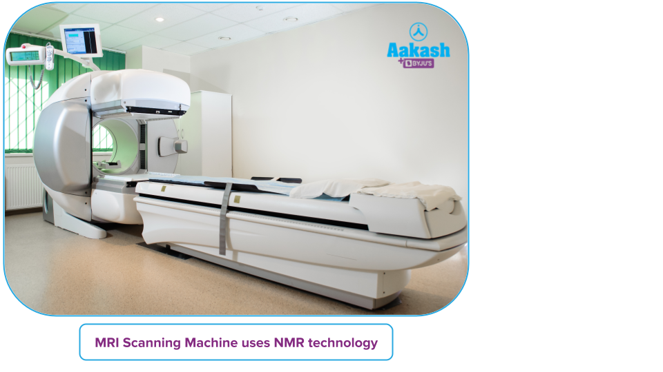
- MRI scan employs NMR technology. For the detection and treatment of illnesses including TB, colon cancer, pneumonia, Parkinson's disease, and many others, biomarkers are a valuable tool.
Practice Problems
Q1. Which region from the electromagnetic spectrum corresponds to NMR?
A. Infrared rays
B. Radio frequency
C. Microwave
D. UV rays
Answer: B)
Solution: In nuclear magnetic resonance (NMR), the radio-frequency zone (3 MHz -30,000 MHz) is where the samples absorb electromagnetic radiation at frequencies determined by the properties of the sample.
So, option B is the correct answer.
Q2. Chemical shift is denoted by the symbol?
A.
B.
C.
D.
Answer: C)
Solution: Chemical shift refers to differences in electron distribution that cause changes in nuclear magnetic resonance frequencies of the same kind of nucleus. It is denoted by the symbol δ.
So, option C is the correct answer.
Q3. What is proton NMR spectroscopy?
Answer: Proton nuclear magnetic resonance is a kind of application of the NMR spectroscopy technique to(Hydrogen-1) nuclei in a substance’s molecules to determine the structure of its molecules. It is also known as Spectroscopy.
Q4. Why is the chemical shift in CH4 lower than CH3Cl?
Answer: Since chlorine is an electronegative element, it will attract electrons to it, deshielding the hydrogen nucleus. The chemical shift (in ppm) will thus be greater in CH3Cl. Since the hydrogen nucleus is protected in the case of CH4, the peak is seen on the lower ppm side.
Frequently Asked Questions – FAQ
Q1. Why are radio waves used in NMR?
Answer: NMR employs a powerful magnet to examine the atomic nuclei's intrinsic spin characteristics. NMR, like other spectroscopies, promotes transitions between nuclear energy levels by using a component of electromagnetic radiation and for enhanced resonance of nuclear energy levels, radio waves which are of high frequency are used.
Q2. What does resonating frequency signify in NMR spectroscopy?
Answer: It corresponds to the absorption energy and the signal's intensity, which are inversely related to the magnetic field's strength. When exposed to a magnetic field, NMR active nuclei absorb electromagnetic radiation at a frequency specific to the isotope.
Q3. How is an MRI scan based on NMR?
Answer: Nuclear magnetic resonance (NMR), the foundation of which is radiofrequency (RF) electromagnetic waves with a particular resonance frequency, is the basis for magnetic resonance imaging (MRI). In Nuclear Magnetic Resonance (NMR) spectroscopy, the identification of an unknown substance (such as a possible new medication) may be determined by the resonant qualities (the jiggling of protons) of the atoms that constitute it. MRI employs a similar physical process.
Q4. How sensitive is NMR spectroscopy?
Answer: Because a measurement using NMR needs a sizable amount of sample, it is an insensitive technology. For instance, although a mass spectrometry experiment only needs roughly 1 μg, an NMR experiment needs between 5-25 mg sample. So the trace amount is difficult to be qualitatively analysed by this technique.