-
Call Now
1800-102-2727


Structure and Function of the Cell Wall and Cell Membrane, Practice Problems and FAQs
Let us compare the cell to our homes. Every home is a safe space surrounded by walls and a roof to keep us protected from the outside world. Did you know that every cell also has a protective cover to keep it safe from the external environment? Now the degree of protection varies depending on the type of cell, just like the number of protective boundaries vary from one home to another. Some houses have an additional boundary wall surrounding the entire house while some don’t. The former is the case of plant cells, fungal cells, algal cells and bacterial cells which have a cell wall as well as a cell membrane protecting their inner contents while the animal cells resemble the latter and are protected only by the cell membrane. In this article, we will discuss the cell wall and the cell membrane of cells and try to understand their structure and function.
Table of Content
Cell wall
The thick porous outer protective covering in bacteria, fungi, algae and plants is known as cell wall. It can provide rigidity and strength to the cells. Another major function of the cell wall is to facilitate cell to cell interaction. Cell walls are absent in animal cells.
- Bacterial cell wall is mainly composed of a polymer named peptidoglycan which is composed of alternate units of N-acetyl glucosamine (NAG) and N-acetyl muramic acid (NAM).

Fig: Structure of peptidoglycan
- The cell wall in fungi has chitin, a polymer of N-acetyl glucosamine (NAG) units, as its major component. It also consists of glucans.

Fig: Fungal cell
- The algal cell wall is composed of cellulose, mannans, galactans and minerals like calcium carbonate.

Fig: Algal cell
- The plant cell wall is primarily composed of cellulose and also contains other complex polysaccharides such as hemicellulose and pectin along with proteins.

Fig: Plant cell
Structure of plant cell wall
The structure of the plant cell wall comprises an extremely intricate lattice and is composed of a matrix and microfibrils. Cell wall depositions are found in the secondary cell wall in plants.
A young plant cell consists of a single primary cell wall but as they stop growing, the secondary walls are formed inside the primary cell wall. The plant cell wall thereby consists of three layers -
- Middle lamella
- Primary cell wall
- Secondary cell wall
Fig: Arrangement of of the different layers of plant cell wall
Middle lamella
It is a thin layer between two adjacent cells which holds them together and is formed in all cells right after cell division. It is composed of calcium pectate and magnesium pectate and can be dissolved by strong acids.

Fig: Middle lamella
Primary cell wall
It is the true cell wall formed in a young plant cell and lies just outside the cell membrane. It is the first formed wall of a plant cell and grows by intussusception. It is around 1-3 μ thick and elastic in nature.
The primary cell wall is formed of an amorphous gel - like matrix in which a loose network of cellulose microfibrils is embedded. The matrix consists of water, pectin, hemicellulose, lipids and glycoproteins in plant cells.
Fig: Primary cell wall
Secondary cell wall
The secondary cell wall is laid down by the cytoplasm inner to the primary cell. It grows by accretion or apposition. It is around 5-10 μ thick and is rigid in nature.
It is usually laid down in three successive layers or more. The cellulose microfibrils in the secondary cell wall are embedded in a gelatinous matrix made up of hemicellulose but the orientation of the microfibrils is different in each layer.
Fig: Layers of secondary cell wall
The secondary cell wall has depositions of various substances such as cutin, suberin, lignin, etc in the wall matrix. These thickening materials are secreted by the protoplasm and may be deposited uniformly or in different patterns.
Fig: Different patterns of secondary cell wall depositions
Pits
The regions where the secondary cell wall is absent are known as the pit. Pits in adjacent cells lie opposite to each other in pit pairs and help in rapid translocation of materials between the cells. The pits on the free surface of the cell are called blind pits as they do not have a partner.
Pits formed of only the primary wall are called simple pits and the ones which are overhung by adjoining thickening matter are called bordered pits.

Fig: Bordered pits
Plasmodesmata
The fine strands of cytoplasm that run through fine pores in the middle lamella and cell walls are called plasmodesmata and help in connecting adjacent plant cells. These facilitate movement of substances between cells.
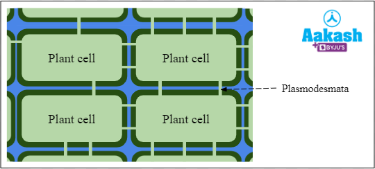
Fig: Plasmodesmata
Functions of Cell Wall
The cell wall performs many functions. Some of them are as follows:
- Helps to maintain the shape and size of the cell and provides mechanical support.
- Protects the cell from osmotic shocks, injuries and pathogens.
- Allows transport of materials in and out of the cell.
- Helps in exchange of materials between adjacent cells through plasmodesmata.
- Helps the cell to enlarge.
Cell Membrane
The membranous covering that surrounds the cytoplasm of all cells is known as the cell membrane. The structure of the cell membrane is the same in both prokaryotes and eukaryotes. It is a semipermeable membrane and crucial in regulating what goes in and out of the cell and also helps the cell to interact with its surroundings.
Structure of cell membrane
The cell membrane is not distinguishable under the light microscope and hence its detailed structure has to be studied under an electron microscope. Through chemical analysis of the cell membrane and detailed study through electron microscopy, scientists were able to determine the structure and composition of the cell membrane. The cell membrane is a trilaminar structure with around 75Å thickness. It has an outer electron dense layer (20 Å), middle pale coloured layer (35 Å) and inner electron dense layer (20 Å). Extended ꞵ proteins form the outer layer and inner layer and phospholipids make up the middle layer.
The major components of a cell membrane are phospholipids, peripheral proteins, integral proteins, transmembrane proteins, cholesterol and carbohydrates attached to the lipids (glycolipids) or proteins (glycoproteins) on the extracellular side of the membrane.

Fig: Structure of Cell membrane
Fluid Mosaic Model of Cell Membrane
Seymour Jonathan Singer and Garth L. Nicolson described the most widely accepted and highly advanced model of the cell membrane structure in early 1975.

Fig: Singer and Nicolson
This fluid mosaic model states that the cell membrane is a quasi-fluid structure in which the phospholipid and protein molecules are arranged in a mosaic in nature. The phospholipids form a bilayer and the proteins are embedded in this bilayer in a mosaic fashion.

GIF: Fluid mosaic model

Fig: Fluid mosaic model of cell membrane
Lipids
The lipids found in the cell membrane are mostly phospholipids which are arranged in two layers (bilayer) with a total thickness of around 35Å. Being ‘amphipathic’ in nature, the polar hydrophilic heads of the phospholipids face the outer sides while the non-polar hydrophobic tails face the inner side.

Fig: Lipid bilayer
Proteins
Proteins in biomembranes are of three types:
- Peripheral proteins that reside on the inner or outer surface of the membrane.
- Integral proteins that are present within the membrane layers.
- Transmembrane proteins that are partly present inside and partly outside the cell membrane.

Fig: Types of membrane proteins
Functions of cell membrane
The quasi fluid structure of the plasma or cell membrane allows the cell to carry out various functions such as endocytosis, cell division, secretion of substances, growth, formation of cell junctions, etc. Some of the functions of the cell membrane are:
- Allows the passage of fat soluble substances across itself.
- Allows the cells to move.
- Facilitates the change of shape of cells for different functions.
- Helps in cell to cell recognition.
- Allows selective transport of molecules and ions through it.
Practice Problems
Q1. What is the difference between growth by intussusception and apposition?
Answer:
|
Growth by intussusception |
Growth by apposition |
|
Growth due to the addition of more wall material from within the existing wall is called intussusception. |
Growth external deposition of new wall material as thin plates on the inner surface of existing wall. |
|
It occurs in the primary cell wall. |
It occurs in the secondary cell wall. |
Q2. Which of the following options correctly represents the trilaminar structure of the cell membrane?
A. outer electron dense layer (20 Å), middle pale coloured layer (35 Å) and inner electron dense layer (20 Å)
B. outer pale coloured layer (35 Å), middle electron dense layer (20 Å) and inner electron dense layer (20 Å)
C. outer electron dense layer (20 Å), middle electron dense layer (20 Å), and inner pale coloured layer (35 Å)
D. outer electron dense layer (35 Å), middle pale coloured layer (20 Å) and inner electron dense layer (35 Å)
Solution: The cell membrane is a trilaminar structure with around 75Å thickness. It has an outer electron dense layer (20 Å), middle pale coloured layer (35 Å) and inner electron dense layer (20 Å). Extended ꞵ proteins form the outer layer and inner layer and phospholipids make up the middle layer. Thus, the correct option is a.
Q3. Which of the following statements is correct?
A. Secondary cell wall is added to the plant cell while it's still growing.
B. The secondary cell wall is laid down outside the middle cell wall.
C. The areas where the secondary cell wall is absent are called pits.
D. All the pits present of the secondary cell wall of a cell are paired pits.
Solution: The secondary cell wall is added to those plant cells that have stopped growing. It is laid down by the protoplasm on the inner side of the primary cell wall. Certain areas in such cells are devoid of the cell wall and such areas are known as pits and help in translocation of materials between adjacent cells. Pits in adjacent cells lie opposite to each other in pit pairs but the pits on the free surface of the cell are called blind pits as they do not have a partner.
Thus, the correct option is c.
Q4. In the phospholipid bilayer
A. the hydrophilic heads are towards the inner side and the hydrophobic tails are towards outer side
B. the hydrophilic tails towards the inner side and the hydrophobic heads are towards outer side
C. the hydrophilic tails are towards the outer side and the hydrophobic heads are towards inner side
D. the hydrophilic heads are towards the outer side and the hydrophobic tails are towards inner side
Solution: The lipids found in the cell membrane are mostly phospholipids which are arranged in two layers (bilayer) with a total thickness of around 35Å. Being ‘amphipathic’ in nature, the polar hydrophilic heads of the phospholipids face the outer sides while the non-polar hydrophobic tails face the inner side. Thus, the correct option is d.
FAQs
Q1. Which type of plant cells consist of only the primary cell wall?
Answer: Meristematic cells, parenchyma cells and the cells in leaves and fruits consist of the primary cell wall only.
Q2. Do plant cells have any tertiary cell wall?
Answer: In tracheids of gymnosperms, an additional cell wall is present on the inner side of the secondary cell wall. It is composed of cellulose microfibrils and xylans.
Q3. What is meant by Robertson’s unit membrane theory?
Answer: Robertson’s unit membrane model proposes that all living biomembranes have the same trilaminar structure in which a lipid layer is sandwiched between two layers of protein.
Q4. What is sarcolemma?
Answer: The cell membrane of the muscle cell is called sarcolemma. It is an excitable membrane similar to the axolemma (membrane surrounding the axon of neurons) in which an action potential can be generated.
YOUTUBE LINK: https://www.youtube.com/watch?v=-NOY_k8iN9A&t=2525s (33:51- 39:35)
https://www.youtube.com/watch?v=-NOY_k8iN9A (20.55- 24.33, 39.37- 42.04)