-
Call Now
1800-102-2727


Regulation of Respiration: Neural and Chemical Regulation, Practice problems and FAQs
In school you might have participated in some activities related to sports like running races, long jumps etc. While taking part in a running race you might have noticed that you breathe fast during running. Why is it happening? While running our body requires more oxygen to produce more energy, to compensate for the additional requirement and hence we breathe fast.
GIF: Running race

We all need energy to carry out various functions in our body, right?. We all know that this energy is coming from the oxidation of food. During the process of oxidation of food oxygen is used to produce energy and carbon dioxide and water are formed as by-products. The carbon dioxide formed is harmful to the body and hence it is removed. This entire process includes breathing and cellular respiration and together it is called respiration.
Now place your hands on your chest and feel what happens? You can feel an upward and downward movement. Yes, you know it happens because of breathing.
GIF: Breathing

Breathing is an involuntary (not under our control) process. It is the process of exchange of oxygen from the atmosphere with carbon dioxide produced by the cells. A normal adult human breaths 12 -16 times/min and infant breaths about 44 times/min.
You know humans have the ability to regulate breathing rate according to the needs of the body. This regulation of respiration involves both neural and chemical control. Now let’s take a deep dive into the article and learn more about the regulation of respiration.
Table of contents
- Regulation of respiration
- Neural regulation
- Chemical regulation
- Practice problems
- FAQs
Regulation of respiration
Human beings have a significant ability to maintain the respiratory rhythm to suit the body needs. This is called the regulation of respiration.
Components involved in regulation of respiration
The regulation of respiration is mainly dependent on the interactions of three components of the respiratory system. These are as follows:
Control centres
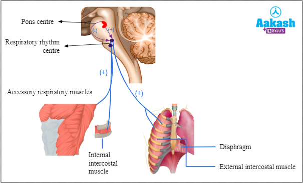
These are present in the brain stem (pons varolii and the medulla oblongata) and are responsible for the automaticity of breathing. Input for these centres comes from the higher brain centres to produce the required voluntary breathing efforts.
Sensors
These include chemoreceptors and sensory receptors.
Chemoreceptors
The chemoreceptors respond to changes in the blood carbon dioxide, oxygen, and hydrogen ion concentration by sending impulses to the control centres. This will alter the breathing pattern by affecting the effector organs.
Sensory receptors
The sensory receptors are located in the upper and lower airways, the lung, and the muscles of respiration.
Effector organs
This includes the respiratory muscles like the diaphragm, the rib cage muscles (external intercostal muscles and internal intercostal muscles) and the abdominal muscles.
Fig: Regulation of regulation
Types of respiratory regulations
There are two types of respiratory regulations as follows:
- Neural regulation
- Chemical regulation
Fig: Regulation of respiration

Neural regulation
The respiratory rhythm is regulated by respiratory centres, which is composed of group neurons located in the hind part of the brain. The rate and depth of breathing is regulated by these respiratory centres. There are two regions in the brain which mainly regulate respiration. These are as follows:
- Medulla oblongata
- Pons varolii
Medulla oblongata
It is located in the hindbrain. It can regulate both expiration and inspiration depending on the neurons activated. It regulates respiratory rhythm according to the requirements of the body. Medulla oblongata has two respiratory centres as follows:
- Dorsal respiratory group (DRG)
- Ventral respiratory group (VRG)
Fig: Respiratory centres of medulla oblongata

Dorsal respiratory group (DRG) or inspiratory group (IG)
It is located in the dorsal portion of the medulla oblongata. This group of neurons mainly causes inspiration by stimulating the muscles of the diaphragm to flatten and the external intercostal muscles to raise the ribs. This group mainly maintains respiratory rhythm and respiratory rate.
As DNG regulates the process of inspiration, it is also called the inspiratory centre or respiratory rhythm centre.
Fig: Dorsal respiratory group (DRG) or inspiratory group (IG)

Ventral respiratory group (VRG) or expiratory group (EG)
It is located in the ventrolateral part of the medulla oblongata. It sends signals for both inspiration and expiration. To regulate inspiration it sends signals to diaphragm and external intercostal muscles. To regulate expiration it sends signals to internal intercostal muscles and muscles of the abdominal wall. It is mainly responsible for regulation of forceful expiration and is also known as expiratory centre.
Fig: Ventral respiratory group (VRG) or expiratory group (EG)

Pons respiratory centres
It is present in the pons varolii of the hindbrain. It regulates the respiratory rhythm centre.
Fig: Pons respiratory centre

There are two respiratory centres present in the pons varolii as follows:
- Pneumotaxic centre
- Apneustic centre
The apneustic centre
This centre lies in the lower part of the pons varolii and sends signals for inspiration for deep and long breaths. It is responsible for exciting the inspiratory centre. Stimulation of this centre results in a gradual increase in the rate of contraction of the inspiratory muscles. It increases tidal volume. It normally works in coordination with the pneumotaxic centre.
Fig: Apneustic centre

The pneumotaxic centre
It is located in the dorsal part of the pons varolii. It can slow down and reduce the functions of the respiratory rhythm centre. Signals from this centre can decrease the duration of inspiration which in turn will alter the respiratory rate. It works through the medullary centres. The pneumotaxic centre sends signals to inhibit inspiration which allows it to specifically control the respiratory rate. It reduces the activity of the phrenic nerve and inhibits the signals of the apneustic centre too. It decreases the overall tidal volume. The apneustic and pneumotaxic centres are antagonistic in function (work against each other together), thereby controlling the respiratory rate.
Fig: Pneumotaxic centre

Hering-Breuer reflex arch
It is a protective reflex as it prevents alveoli from bursting. It is initiated to prevent the over-inflation of the lungs. There are stretch receptors present in the walls of terminal bronchioles and atria. When maximum air gets filled in alveoli, these receptors are activated. This in turn activates the Hering Breuer Reflex Arch which sends inhibitory signals to the inspiratory centre through the vagus nerve to switch off the inspiration and expiration begins. This prevents alveoli from overstretching or bursting.
Fig: Hering-Breuer Reflex Arch

Chemical regulation
Centres associated with chemical regulation of respiration are as follows:
- Central chemoreceptors
- Peripheral chemoreceptors
Central chemoreceptors
These are present in the medullary oblongata region around the inspiratory centre. This area is sensitive to pCO2 and H+ ions concentration. Hence an increase in pCO2 and H+ ions activate these receptors, which in turn activate respiratory rhythm centre or the inspiratory centre. Activated rhythm centres alter the rate of respiration or inspiration. This will make necessary adjustments in the respiratory process by which these substances will be eliminated.
Fig: Chemical regulation of respiration

Peripheral chemoreceptors
These include the following:
- Aortic bodies
- Carotid bodies
These are activated by increase in pCO2 and H+ ions in arterial blood. These activate the inspiratory centre or respiratory rhythm centre to reverse the situation.
Aortic bodies
It includes the chemoreceptors present in the arch of aorta. Their afferent nerve fibres pass through the vagus nerve ( tenth cranial nerve) to the dorsal respiratory group.
Carotid bodies
It includes the large number of chemoreceptors present in the bifurcations of the common carotid arteries. Their afferent nerve fibres pass through the glossopharyngeal cranial nerves and reach the dorsal respiratory group of neurons in the medulla oblongata.
Fig: Chemical regulation

Practice Problems
- Which regions of the brain help to regulate respiration?
- Pons varolii
- Medulla oblongata
- Cerebellum
- Both a and b
Solution: The respiratory rhythm is regulated by respiratory centres, which is composed of group neurons located in the pons varolii and medulla oblongata. The rate and depth of breathing is regulated by respiratory centres. There are two main respiratory centres in the brain as follows:
- Medullary respiratory centres (Respiratory rhythm centre)
- Pons respiratory centres (Pneumotaxic centre and apneustic centre). Hence, the correct option is d.
2. Which of the following prevents the lungs from expanding too much during inspiration?
- Medullary centre
- Pneumotaxic centre
- Apneustic centre
- All of the above
Solution: Pneumotaxic centres can slow down and reduce the functions of the respiratory rhythm centre. Signals from this centre can decrease the duration of inspiration which in turn will alter the respiratory rate. It works through the medullary centres. The pneumotaxic centre sends signals to inhibit inspiration which allows it to specifically control the respiratory rate. Hence, the correct option is b.
3. Identify the site of the pneumotaxic centre?
- Medulla oblongata
- Pons varolii
- Cerebrum
- Cerebellum
Solution: The pneumotaxic centre is located in the pons varolii of the brain. Pneumotaxic centres can slow down and reduce the functions of the respiratory rhythm centre. Signals from this centre can decrease the duration of inspiration which in turn will alter the respiratory rate. Hence, the correct option is b.
4. Overstretching of lungs can be prevented by ___________.
- Bohr’s effect
- Haldane’s effect
- Herring-Breuer reflex
- All of the above
Solution: Herring-Breuer reflex is a protective reflex as it prevents alveoli from bursting. It is initiated to prevent the over-inflation of the lungs. There are stretch receptors present in the walls of terminal bronchioles and atria. When maximum air gets filled in the alveoli, these receptors are activated. This in turn activates the Hering Breuer Reflex Arch which sends inhibitory signals to the inspiratory centre through the vagus nerve to switch off the inspiration. This prevents alveoli from overstretching or bursting. Hence, the correct option is c.
FAQs
- What is the basic mechanism involved in respiration?
Answer: The respiration involves the breathing mechanism and exchange of gases. The breathing involves the inspiration of oxygen and expiration of carbon dioxide. The exchange of gases occurs at the alveoli through a process of diffusion.
- Which nerves are responsible for controlling the phenomenon of breathing?
Answer: The phrenic nerves control breathing. These nerves allow the diaphragm to contract and expand which allows the lungs to inhale and exhale air.
- Which muscles are responsible for respiration?
Answer: There are three groups of respiratory muscles that are involved in respiration. These are as follows:
- Diaphragm
- Rib cage muscles (External intercostal muscles and internal intercostal muscles)
- Abdominal muscles
4. Which is considered as the main regulator of respiration?
Answer: Medulla oblongata functions as a relay station between the spinal cord and the brain. It contains centres for regulating vasomotor, cardiac, respiratory and reflex activities.
Related Topics
|
Introduction to Respiration, Respiratory organs in animals, Practice Problems and FAQs |
|
Lung Volumes and Capacities, Practice Problems and FAQs |
|
Mechanism of breathing, Difference between inspiration and expiration, Abdominal breathing, Thoracic breathing, Hering - Breuer reflex, Practice Problems and FAQs |
|
Transport of gases and oxygen dissociation curve |
|
Exchange of gases: Partial pressure, Diffusion, Sites of exchange of gases, Practice Problems and FAQs |