-
Call Now
1800-102-2727


Sense Organs: Eyes, Ears, Tongue, Nose, and Skin, Practice Problems, and FAQs
How many times in our lives have we touched a cup of hot tea or coffee and immediately regretted it? The moment we touch something extremely hot, we immediately pull our hands away. Even when we feel an abrupt and unexpected touch, we react impulsively. These reactions are due to reflex actions that occur immediately after the skin receives the stimulus of heat or an abrupt touch. Reflex actions such as these can often help us to defend ourselves from dangers around us. The skin that covers the whole body constitutes 16% of the total body mass and is considered the largest organ of the human body. It has many receptors that receive a variety of different stimuli and send information to the brain or the spinal cord to be processed accordingly.
How do you feel when your nose is blocked during a cold and cough? Have you noticed that we often lose the appetite to eat at such times? One of the major reasons behind this is that we are not able to smell or taste our food properly. Do you know why? This is because the mucus in the nasal cavity blocks the smell sensing receptors present in our nose. As the sense of smell and the sense of taste together help us in perceiving the flavour of food in its entirety, the impairment of sensory receptors in the nose prevents us from developing an appetite for food. This is more prominent in pets like cats and dogs who entirely reject food during a cold as they can’t smell it.
Try blind folding yourself for a few hours and try to do all the chores that you would normally do in a day. Pretty sure that you would find it extremely difficult to accomplish even the simplest of tasks if you are not able to see anything around you. This puts us in the perspective of how difficult it must be for visually challenged people. However, they are more competent in getting through their day even without the sense of sight because they acquire reflexes through daily habits and routines.
Thus, it is fairly justified to say that to be able to sense stimuli around us is very crucial for our survival. And what helps us in sensing stimuli? The sense organs and the sensory receptors in our body. Let’s dive into this article and study the different types of sense organs and their receptors.

Table of contents
- Sensory receptors
- Mechanism of Sensory reception
- Properties of receptors
- Sense organs
- Eyes
- Ears
- Tongue
- Nose
- Skin
- Practice Problems
- FAQs
Sensory receptors
A sensory receptor is either a neuron ending or a specialised cell that is in close contact with a neuron ending which produces a nerve impulse in response to a stimulation in the internal or external environment. The nerve impulse then travels to the central nervous system via the sensory (afferent) neurons for processing and response formation.
The senses perceived by the receptors are categorised into two types:
- Special senses - These types of senses have specialised receptors that gather sensory information from the interacting environment and change it into nerve impulses. Examples of special senses include vision (eyes are specialised sense organs), hearing (ears), balance (ears), taste (tongue), and smell (nose).
- General senses - In contrast to special senses, general senses are associated with the sense of touch. They do not have special sensory receptors.
Sensory receptors can be classified based on their position:
Exteroreceptors
These receptors are found at or close to the body surface and receive external stimuli such as heat, light, pressure, pain, etc. Exteroreceptors can be of the following types -
General receptors
These receptors sense the general sense of touch and are present on the skin. These are also known as cutaneous receptors. Mechanoreceptors which respond to various stimuli of mechanical forces, such as pressure, roughness, vibrations, and stretching are examples of such cutaneous receptors.
Thermoreceptors on the skin that can respond to variations in temperature and sense temperatures above or below body temperature are also general receptors.
Specialised receptors
These include the photoreceptors or light receptors in the eyes, olfactory receptors in the nose, audio receptors in the ears and the tangoreceptors in the tongue. The receptors in the nose and tongue are specialised chemoreceptors that respond to specific chemicals present in the air or food, respectively. Nocireceptors on the skin are specialised receptors that respond to potentially damaging stimuli, such as pain, damaging heat or cold, excessive pressure, or painful chemicals.
Interoceptors or Visceral receptors
These receptors are present in the walls of blood vessels and different internal organs. These can be nocireceptors, thermoreceptors, chemoreceptors or mechanoreceptors. These occur as -
- Naked nerve terminals in the visceral walls or outer fibrous layer of blood vessels
- Lamellated corpuscles in heart, pancreas, mesenteries and fibrous outer layer of blood vessels
- Arborised terminals in endocardium of heart, endomysium of muscles and in connective tissue
- Baroreceptors or pressure receptors in walls of blood vessels, alveoli, etc.
Proprioceptors
The conscious or unconscious sense of joint position is referred to as proprioception. This system aids the body with locating the joints, muscles, and limbs in three dimensions as well as the direction in which each is moving with respect to the rest of the body. Examples of the proprioception system include walking or kicking without looking at our feet, balancing on one leg, touching the nose with our eyes closed, and the ability to sense the surface on which we are standing upon. These receptors are mostly found in the muscles, tendons, joints, etc.
Mechanism of Sensory reception
Receptor potentials are the starting point of all sensory impulses. These potentials cause a neurotransmitter to be released, which then stimulates the relevant neuron to transmit information to the brain. In order to generate a receptor potential, the action potential of the receptor cell membrane must rise above a threshold level, just like with conventional neuron signal transduction. The generation of the action potential is triggered by the stimuli received. As the threshold value of sensory receptors increases, the frequency of action potentials also increases. All receptors have the ability to recognise both strong and weak signals. When the stimulus reaches its maximal level of excitation, there is a drop-off or plateau. The receptor is then unable to boost its firing potential.
The impulses are carried to the brain via the sensory neurons. Hypothalamus is the main centre and the cerebral cortex is the second centre for the processing of the sensory impulses in the brain.
Properties of receptors
There are some properties that are common in all types of sensory receptors. These are discussed below:
- Receptive field
- Labelled line principle
- Adaptation
Receptive field
The receptive field is described as the area of a body where a stimulus can affect the sensory receptors. To accurately encode the location of a stimulus, this property is crucial in the form of a physical dimension. The fovea of the retina and regions of the skin like the fingertips and lips are examples of areas with a higher density of tiny receptor fields that are capable of achieving enhanced spatial resolution.
Labelled line principle
The sensory system works by reacting to the stimuli they are specially designed for and then translating this stimulus into the neuronal message that travels through a discrete path to the brain. This is termed a labelled line principle that reserves the specificity of a receptor class in encoding a sensory message which is specific to the designated brain area. This principle is applied to the somatosensory systems as well as specialised systems, such as auditory and visual.
Adaptation
It is a very common property of all types of sensory receptors. There will be a decline in the rate of action potentials as a stimulus repeatedly excites the receptor. Receptors are able to adapt to the change in the stimulus or intensity.
Sense organs
Sense organs are the multicellular organs formed of more than one sensory receptor. They receive the environmental stimuli and transmit these impulses to the central nervous system.
Sense organs are described as the specialised organs that help an organism in perceiving the surrounding environment. They are a necessary part of our existence and the only means by which we can understand our surroundings. Sense organs transfer the signals from the reactions taking place in the surrounding environment with the help of a network of nerves. The sense organs help to regulate the associations and interactions between the human body and the environment. There are five sensory organs in the human body, that are listed below:
- Eyes
- Ears
- Nose
- Tongue
- Skin
These five sense organs have receptors that function to receive information from the surroundings and send them to the relevant part of the nervous system with the help of sensory neurons.
Eyes

Fig: Eye
The eyes are known as our body’s visual sense organs and therefore, they are used for vision or ophthalmoception. These organs are sensitive to light images and are like hollow balls which are located in a bony socket called ‘orbit’ in the skull. The wall of the eyeball is made up of three layers – outermost fibrous coat, middle vascular coat and innermost retina.
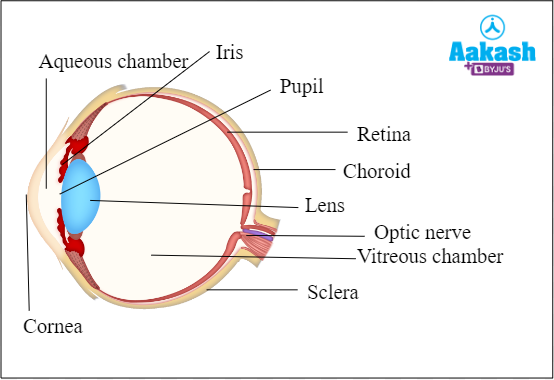
Fig: Eyeball
The outermost layer of the eyeball is made up of two parts - the white sclera composed of fibrous connective tissue and the bulged and transparent cornea in the front.
The middle layer of the eyeball contains the melanin pigment and is richly supplied with blood vessels. It is divided into three parts - choroid, ciliary body and iris. The choroid is the melanin rich layer which prevents internal reflection of light rays to avoid blurring of the image and helps to produce a sharp image.
The iris is an opaque but muscular pigmented structure which determines the colour of the eye. It is a perforated diaphragm that has a central perforation known as the pupil in the anterior part of the middle layer. The iris muscles contract and relax to dilate and constrict the size of the pupil, respectively. This helps to control the amount of light entering the eye.
The ciliary body is a thick vascular, less pigmented, ring shaped structure that lies at the junction of iris and choroid. It has folds on the inner side known as ciliary processes. Suspensory ligaments are attached to the lens on one end and the ciliary body on the other end. The ciliary processes and the suspensory ligaments together help in changing the shape of the lens to focus at different distances.
Retina is the innermost layer of the eyeball which has sensory receptors and is non vascular in nature. The retina has two parts - an outer pigmented part and inner nervous layer. The inner sensitive part of retina is made up of three layers -
- Outer layer consisting of photoreceptor cells of two types - rods and cones. It also has modified unipolar nerve cells.
- Middle layer of bipolar nerve cells.
- Inner layer of ganglion cells

Fig: The layers of retina
Rod cells
These types of cells are present at the margin of the retina and they help in vision in darkness or dim light, i.e, scotopic vision.. They are also useful for peripheral vision. The rod cells produce the rhodopsin pigment which helps in vision in low light conditions.

Fig: Rod cells of retina
Cone cells
These types of cells are helpful in vision in bright light and colour detection. The cone cells are of three different types based on their ability to detect the major colours of light. These are blue, green, and red cells. Cone cells produce a photosensitive pigment called iodopsin.
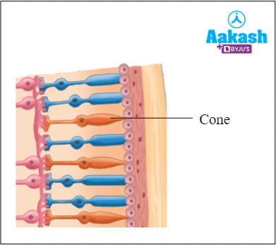
Fig: Cone cells of retina
The eye lens is the transparent, elastic, biconvex structure which is suspended behind the pupil, in the cavity of the eyeball, with the help of the suspensory ligaments. It is mainly responsible for refracting the light rays to create an image. The chamber between the cornea and the lens is the aqueous chamber and is filled with a watery substance called aqueous humour. The chamber between the lens and the retina is called the vitreous chamber and is filled with a jelly-like vitreous humour. The aqueous and vitreous humour, along with the lens and cornea also help in refraction of light. These fluids also help in maintaining the shape of the eyeball.
As the light rays form an image on the retina, the light impulses received by the rod or cone cells generate potentials or impulses in them. These impulses are passed from the retina to the visual cortex of the brain through the optic nerve.
Ears
Ears are our auditory sense organs and therefore, they are used for hearing or audioception. They help in the perception of sounds. Our auditory system senses air vibrations and uses this information to create sound. This is known as hearing or audio captioning. An ear is composed of three parts: the outer ear, the middle ear, and the inner ear. The outer ear sends the vibrations into the inner ear which contains specific regions of sensory receptors that convert the vibrations to impulses and send them to the brain via the auditory nerve. The brain converts them into audible sounds because all sounds are essentially vibrations. Apart from hearing, the ears play a crucial role in maintaining the balance of the body or equilibrium.

Fig: Ear
Outer ear
The outer ear is composed of the auricle or pinna (visible portion), a short external auditory canal, and a tympanic membrane or ear drum. Sound waves are gathered by the outer ear and sent to the tympanic membrane which starts vibrating upon receiving the waves. Thus, it converts the waves into mechanical vibrations and passes it on to the middle ear via the first ear ossicle, that is, malleus attached to it.

Fig: External ear
Middle ear
The middle ear is described as a narrow and air-filled cavity present in the temporal bone. It holds three tiny bones named hammer (malleus), anvil (incus), and stirrup (stapes) which are collectively called the ear ossicles. The ear ossicles amplify the sound waves by a lever-like action. The vibrations from the middle ear to the inner ear are transferred by a round window and an oval window.
The eustachian tube connects the middle ear to the pharynx and helps to equalise the air pressure on either side of the eardrum to allow it to vibrate freely.
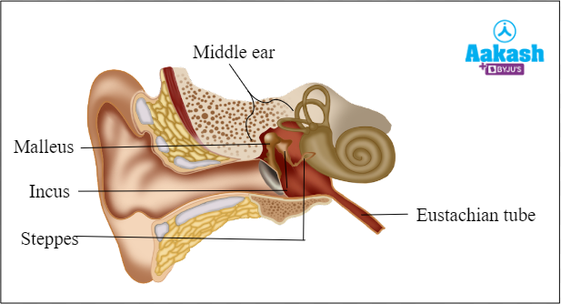
Fig: Middle Ear
Inner ear
The inner ear is composed of a membranous labyrinth present inside the bony labyrinth. A fluid known as perilymph surrounds the bony labyrinth and a fluid named endolymph fills the membranous labyrinth.
In the inner ear, two functional units are present and these are the vestibular apparatus and cochlea. The vestibular apparatus have vestibule and semicircular canals which have sensory receptors that help in maintenance of body equilibrium, whereas the cochlea has audio receptors that help in hearing.

Fig: Inner ear
Vestibular system
The vestibule possesses two chambers, utriculus and sacculus, which have a sensory spot named macula possessing sensory cells. The semicircular canals have sensory cells at the swollen ends of each of the three perpendicularly arranged canals. This region is known as the ampulla.
Any movement in the body, when at rest or during motion causes the endolymph in the inner ear to move and stimulate sensory receptors to generate an impulse. These impulses regarding body motions, head position, and spatial orientation are sent to the brain via the vestibular branch of the auditory nerve.
The system is also involved with the motor functions and helps in various functions, such as
- Maintaining the body posture
- Maintaining the balance of the body
- Stabilise head and body during movement
- Recognising how the bodies are positioned and oriented in reference to the environment
Therefore, the vestibular system performs a crucial role for normal movement and equilibrium. The utriculus and sacculus help in maintaining static equilibrium whereas the semicircular canals help in maintaining dynamic balance.

GIF: Vestibular system responsible for balance
Cochlea
The cochlea is a coiled portion of the membranous labyrinth, which looks like a snail. It is made up of three canals, upper vestibular canal or scala vestibuli, middle cochlear duct or scala media and the lower tympanic canal or scala tympani. The scala media is filled with endolymph and possesses a sensory region called the organ of Corti. The sound vibrations received from the middle ear cause the endolymph to vibrate which stimulates the sensory receptor cells (audio receptors) in the organ of Corti and an impulse is generated which is carried to the brain via the cochlear branch of the auditory nerve.
Tongue
The tongue helps in the perception of different tastes and flavours and therefore, they are used for gustaoception. On the tongue, taste buds are situated between the papillae and help with flavour perception. The sensations of taste and smell frequently work together. It is impossible to taste something entirely if you can't smell it. The sensation of taste is known as gustoception. Chemoreceptors on the tongue perform the same function as those in the nasal cavity. There are four distinct types of taste buds present on the tongue and each of them can detect different types of taste, such as sweetness, sourness, bitterness, and saltiness.
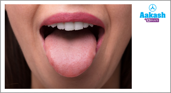
Fig: Tongue
Nose
The nose is our olfactory sense organ used for smell or olfalcoception. The olfactory system helps in detecting different odours. Another term used for the sense of smell is olfaction. The olfactory cells are present on the roof of the nasal cavity. Olfactory cells have olfactory nerve fibres on one end and cilia that extend into the nasal cavity on the other. The air enters the nasal cavity when a person breathes in. Olfactory cells have protein receptors that can detect minute changes in chemicals and are also known as chemoreceptors. These chemicals bind to the cilia and then the sensory information is transmitted to the brain. The brain then converts these impulses into an interpretable fragrance. The body produces mucus during a cold, which suppresses the sense of smell and causes our meals to taste monotonous.

Fig: Olfactory region of nose
Skin
Skin is considered the biggest organ in the human body and is used for touch or tactioception. It aids in perceiving the sensation of touch. The sensory receptors are present in the skin that detect touch, pain, pressure and temperature. An impulse that is produced when skin receptors are active travels from the skin to the spinal cord, then to the brain.

Fig: Skin
Practice Problems
- What are the differences between general senses and special senses?
Answer:
|
General senses |
Special senses |
|
General senses are associated with the sense of touch. They do not need special receptors to be perceived. |
These types of senses have specialised sensory receptors that gather sensory information from the interacting environment and change it into nerve impulses. |
|
Examples include the sense of touch. |
Examples of special senses include vision (eyes are specialised sense organs), hearing (ears), balance (ears), taste (tongue), and smell (nose). |
- Why can skin detect different types of senses, such as pressure and temperature?
Answer: The sense of touch is controlled by a huge network of nerve endings and touch receptors that are present in the skin and they are collectively known as the somatosensory system. These nerve endings rapidly sense the stimulus, such as cold, heat, pressure, pain and many more. These nerve endings then transmit these signals to the specific area of the brain and the brain then interpret the stimulus and react accordingly.
- Identify the function of cone cells in the eyes.
- Secretion and equilibrium
- Vision in dark light
- Monocular vision
- Colour differentiation
Solution: Cone cells present in the retina of our eyes are used in detecting the colours in strong light. The cone cells are of three different types based on their ability to detect the major colours of light. These are blue, green, and red cells. Hence, the correct option is d.
4. Name any five categories of somatosensory receptors.
Answer: The somatosensory receptors are categorised into five categories that are listed below:
- Mechanoreceptors: These types of cells help in detecting mechanical pressure and are found in skin, joints, muscles, tendons, etc.
- Thermoreceptors: These receptors respond to variations in temperature. They can sense temperatures above or below body temperature and are primarily present in the skin.
- Nociceptors: These receptors generally respond to potentially damaging stimuli, such as pain. They are present in internal organs as well as on the outer surface of the body. They detect various stimuli, such as damaging heat or cold, excessive pressure, or painful chemicals.
- Photoreceptors: These receptors detect and respond to light. These receptors are commonly found in the eyes and are required for the sense of vision.
- Chemoreceptors: These receptors are specialised in responding to certain chemicals. They are commonly found in the taste buds as well as in nasal passages.
FAQs
- What will happen if the somatosensory system of a person gets damaged?
Answer: Damage to the somatosensory system can lead to numbness or in some cases, it can also result in paraesthesia. It is a tingling sensation that occurs in certain areas of the body. The damage to the cerebral cortex can result in numbness which then affects the receptors of certain areas.
- What is deafness?
Answer: Deafness is described as the condition that develops when a person loses all or part of their hearing. One or both ears may be affected by this illness.
- Are efferent or afferent nerves used to transmit sensory information to the central nervous system?
Answer: The CNS receives impulses from peripheral organs through the afferent or sensory division. Impulses from the CNS are sent out to the peripheral organs through the efferent or motor division to produce an effect or action.
- Which part of the brain receives the auditory information?
Answer: The auditory sensory information is received by the temporal lobe of the brain.
YOUTUBE LINK: https://www.youtube.com/watch?v=g4FmdE629VA