-
Call Now
1800-102-2727


Anatomy of Monocotyledonous Leaf, Practice problems and FAQs
There is no wonder if you find every monocot leaf alike. This is because most of the monocot leaves are narrow with pointed tips and show the same pattern of the parallel venation. But what do you think about their internal structure? Will it be similar for all the monocot leaves? Do the monocot leaves show any anatomical similarities with the dicot leaves? If yes, then what are their anatomical differences? Are there any specialised structures which are unique to monocot leaves? Have you ever thought why monocot leaves are always more erect than the dicot leaves?
All these answers can be found through analysing the anatomy of a monocot leaf. Observing the transverse section of a monocot leaf under the microscope serves the purpose. It is such beauty to observe the plant cells through a microscope after staining. Let us now take a look at what a monocot leaf looks like under a microscope.
Table of contents:
- Anatomy of monocotyledonous leaf
- Diagram of transverse section of monocot leaf
- Differences between dicot leaf and monocot leaf
- Practice Problems
- FAQs
Anatomy of Monocotyledonous Leaf
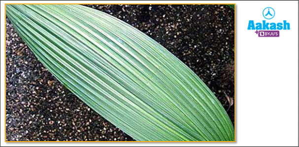
Leaves of plants bearing seeds with a single cotyledon are known as monocotyledonous leaves. Externally, all monocot leaves have parallel venation. They are also called isobilateral leaves as their leaf surfaces are similar on both sides. The anatomy of monocot leaves have some similarities with the dicot leaves, but there are many differences too.
Fig: Monocot leaf
Observing the transverse section of a monocot leaf under the microscope gives us an idea of its anatomical structure. The following structures pertaining to the different plant tissue systems can be observed in the T.S of a monocot leaf -
Epidermal tissue system
The outermost layer or covering of the plant body is formed of the epidermal tissue system. The major function of the epidermal layers is the protection of the leaves. Epidermal tissue system includes the epidermis, stomata and bulliform cells.
Epidermis is single-layer on both the upper (adaxial) and lower (abaxial) surfaces and there is no difference between the two surfaces. The epidermis is made up of compact cells which lack chloroplasts.Outer surface of the epidermis is covered by a water-proof layer of cuticle.
Stomata is present on the epidermis of both sides. So the monocot leaf is called an amphistomatic leaf. Dumbbell-shaped guard cells surround the stomata.
Fig: Monocot stomata

Bulliform cells are large, empty and colourless epidermal cells that are present along the veins on the upper epidermis of grasses. They help in rolling and unrolling of leaves due to change in turgidity. Rolling up of the leaves helps in minimising water loss by reducing the amount of exposed leaf surface area.
Fig: Bulliform cells

Ground tissue system
The ground tissue system in a monocot leaf includes the tissues present between the upper and lower epidermis except the vascular tissues. The only tissue present in ground tissue of monocot leaf is mesophyll.
Mesophyll is present between the upper and lower epidermis and is not differentiated into palisade and spongy parenchyma. They are made up of chlorenchyma (chlorophyll containing parenchyma) cells and form the photosynthetic tissue of the leaf.
Fig: Ground tissue of monocot leaf

Vascular tissue system
The vascular tissue system includes a large number of small, closely placed vascular bundles and a few large vascular bundles in the mesophyll layer of the leaf.
Fig: Vascular tissue

Due to the presence of parallel venation, all the vascular bundles have similar size, except for the ones near the main veins which are larger in size. The vascular bundles are conjoint, collateral and closed. In conjoint vascular bundles, the xylem and phloem combine into a single bundle. In these vascular bundles, if the xylem and phloem lie on the same radius, with the xylem on the inside and phloem on the external side, then it is known as collateral vascular bundles. As the cambium is absent in the vascular bundles, they are known as closed vascular bundles.
Xylem lies towards the upper surface and phloem towards the lower surface of the leaf. Vascular bundles are surrounded by concentric layers of undifferentiated mesophyll cells known as the bundle sheath cells. The bundle sheath cells have larger and higher number of chloroplasts compared to the mesophyll cells. This arrangement of the mesophyll cells around the vascular bundles, having dimorphic chloroplasts, is known as Kranz anatomy.
Diagram of transverse section of monocot leaf
Fig: T.S of monocot leaf

Differences between Dicot leaf and monocot leaf
Features |
Dicot leaf |
Monocot leaf |
Stomata |
More on lower surface |
Equally distributed on both surfaces |
Shape of guard cells |
Bean shaped |
Dumbbell shaped |
Distribution of stomata |
Hypostomatic (stomata present only on the lower side of the leaf) |
Amphistomatic (leaves having stomata on both sides) |
Mesophyll |
Differentiated into palisade and spongy parenchyma |
Undifferentiated |
Size of vascular bundle |
Size of vascular bundles depends on the size of veins which are dissimilar in reticulate venation. Hence, sizes of vascular bundles are also dissimilar. |
Size is similar due to similar size of veins in parallel venation |
Bulliform cells |
Absent |
Present |
Venation |
Reticulate |
Parallel |
Practice Problems
Q 1. Leaves of which of the following plants lacks the palisade parenchyma?
a. Mustard
b. Gram
c. soya bean
d. maize
Answer: Dicot plants include mustard, gram, and soya bean. The mesophyll layer of dicot or dorsiventral leaves is differentiated into palisade and spongy parenchyma. Palisade parenchyma is made up of vertically oriented, parallel-to-each-other elongated cells. They can be found in the adaxial epidermis. The cells in spongy parenchyma are oval or spherical, with many intercellular gaps and air voids between them. It's found beneath the palisade parenchyma cells.
Maize is a monocot plant, which means its seeds have a single cotyledon or seed leaf. In monocots, unlike dicots, the mesophyll of the leaf is not divided into palisade and spongy parenchyma.
Hence the correct option is d.
Q 2. The internal structure of monocot leaf is similar to dicot leaf in the
a. absence of mesophyll
b. arrangement of stomata
c. absence of stomata
d. presence of stomata
Answer: Both dicot and monocot leaves have stomata in their epidermis. Two guard cells surround a stomatal pore in each stoma. Gaseous exchange and transpiration are aided by stomata. The arrangement of stomata in dicot and monocot leaves, however, differs. The lower epidermis of dicot leaves has more stomata than the upper epidermis. Stomata are evenly distributed in both the upper and lower epidermis of monocot leaves. Mesophyll is the parenchymatous ground tissue that aids photosynthesis in both dicot and monocot leaves.
Hence the correct option is d.
Q 3. Choose the correct statement with reference to the internal structure of leaves.
a. On both surfaces of their leaves, all dicot leaves have an equal number of stomata.
b. In leaves, there are conjoint vascular bundles.
c. The abaxial epidermis is the upper epidermis, whereas the adaxial epidermis is the lower epidermis.
d. Cambium is found in leaves.
Answer: In the leaf, vascular bundles consist of xylem and phloem lying along the same radius and are known as conjoint vascular bundles. The lower epidermis of a dicot leaf has more stomata than the upper epidermis. The adaxial epidermis is the upper epidermis of the leaf, whereas the abaxial epidermis is the lower epidermis. There is no secondary growth in leaves because they lack cambium.
Hence the correct option is b.
Q 4. What are bulliform cells?
Answer: The epidermal tissue system includes bulliform cells. Bulliform cells are huge, colourless, and empty cells on the top layer of the epidermis of grasses that lie close to the veins. They result in rolling up of the leaves when they become flaccid. This helps in reducing water loss by decreasing the exposed surface area of the leaf. The leaves unroll when the bulliform cells are turgid.
FAQs
Q 1. Why are there the same number of stomata on both sides of monocot plant leaves?
Answer: Monocotyledons have leaves that are held vertically and parallel to the sun. As a result, both surfaces of monocotyledons get equal amounts of sunlight, resulting in an equal distribution of stomata on both surfaces.
Q 2. Is Kranz anatomy observed in C4 plants?
Answer: Yes, Kranz anatomy can be observed in the C4 plants like maize and sugarcane. The Kranz anatomy is a particular type of structure found in C4 plants. Here, the vascular bundles are surrounded by a ring-like grouping of mesophyll cells called the bundle sheath cells. The bundle-sheath cells have a greater number of chloroplasts than the mesophyll cells and have a thick wall that is impervious to gaseous exchange. Thus, these cells cannot take up atmospheric carbon dioxide and fix carbon dioxide for photosynthesis via the C4 pathway.
In this pathway carbon dioxide is first fixed in the mesophyll cells in the form of malic acid or aspartic acid which are then transported to the bundle sheath cells and decarboxylated to release carbon dioxide. This carbon dioxide is then fixed by the bundle sheath cells using the RuBisCo enzyme via the C3 cycle. Kranz anatomy's main purpose is to offer a location where CO2 can be concentrated around RuBisCO, preventing photorespiration.
Q 3. What is the significance of monocots in agriculture?
Answer: Monocots are found all over the world and make up the vast bulk of agricultural plants in terms of biomass production. This group contains around 50,000 to 60,000 species. Most of the grains that are cultivated as staple food crops all over the world are monocots. Monocots are not only significant ecologically, financially, and as a food staple, but they also have aesthetic significance.
Q 4. Where does maximum growth occur in monocot leaves?
Answer: In monocot leaves, the organisation of the growth processes is considered as spatially regulated. The actively dividing cells which contribute to growth are present at the leaf's base, growing cells in the middle, and mature cells at the tip. Thus, maximum growth occurs at the leaf base.
YOUTUBE LINK: https://www.youtube.com/watch?v=Vrg9wf5gQd8
Related Topics
|
Epidermal tissue system: Epidermis, Stomata, Epidermal appendages |
|
Tissue system: Ground tissue system |
|
Tissue system: Vascular tissue system |
|
Anatomy of dicotyledonous leaves |