-
Call Now
1800-102-2727


Alimentary canal: Internal anatomy, Histology, Practice Problems and FAQs
You all like to enjoy a variety of food. Some like ice creams, some like biryani or kebabs. When we go to parties we all enjoy the various cuisines.

Fig: Food
But Have you ever thought about how much time it takes this food to get digested? How much time does it take the undigested food to get rid of the body? What changes happen to the body in the digestive system?
The food travels from mouth to the anus for digestion, absorption, assimilation and egestion processes. This passage followed by food is known as the digestive tract or alimentary canal. In this tract there are several stations where food stays for some time. Every station is specialised for a particular function. Let’s understand more about the different parts of the digestive system in this article.
Table of contents:
- Alimentary canal
- Mouth
- Pharynx
- Esophagus
- Stomach
- Small intestine
- Large intestine
- Histology of digestive system
- Practice Problems
- FAQs
The alimentary canal
The alimentary canal or the gastrointestinal tract is a long, hollow, coiled, muscular, and glandular tube of variable diameter. The length of the alimentary canal is about 6 - 9 meters. The mouth forms the anterior opening of the digestive tract and anus forms the posterior opening. It contains a number of specialised sections. These sections perform various specialised functions and help in digesting, absorbing and assimilating the food. The digestive system consists of the alimentary canal and the accessory digestive glands. The various parts of the alimentary canal are listed below:
- Mouth
- Buccal cavity
- Pharynx
- Oesophagus
- Stomach
- Small intestine
- Large intestine
- Anus
The various accessory digestive glands are enlisted below:
- Salivary glands
- Gastric glands
- Liver
- Pancreas
- Intestinal glands
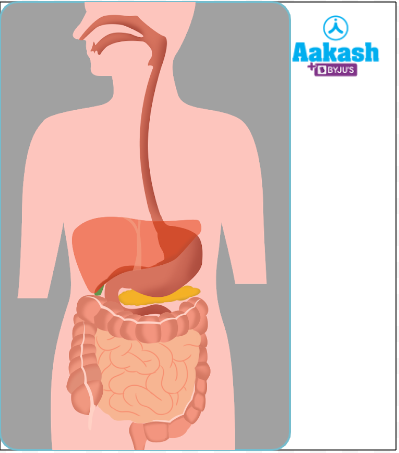
Fig: Alimentary canal
Mouth
Mouth is the first specialised organ of the alimentary canal. It is protected by the upper lip and lower lip. Both the lips are covered with skin on the outer side and lined with mucous membrane on the inner side. The mouth leads to the vestibule, it is a narrow space. Vestibule leads to the next region called buccal cavity.
Fig: Mouth
Buccal cavity
It is located inner to the mouth and is composed of three parts as follows:
- Palate
- Tongue
- Teeth
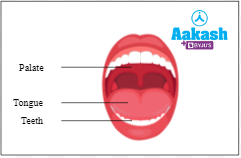
Fig: Parts of buccal cavity
Palate
The roof of the buccal cavity is considered the palate. Rugae are ridges that can be found on the palate. The palate is divided into three sections:
Hard palate
It is the anterior part of the palate that possesses ridges. It helps in chewing.
Soft palate
It forms the posterior part of the palate and is smooth and fleshy.
Uvula
It is considered as the extension of the soft palate and is found hanging above the throat. It helps in swallowing the food particles. It also prevents the entry of food into the nasal chamber.
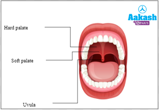
Fig: Parts of a palate
Tongue
The tongue is a flexible, muscular organ that sits on the floor of the mouth. It is a voluntary flat organ. The tongue has sensory functions and aids in the mastication of food. The tongue is divided into three regions, an anterior oral part, posterior pharyngeal part and an inverted V-shaped furrow called sulcus terminalis. A median pit called foramen- caecum is present.
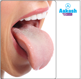
Fig: Tongue
Parts of tongue
Tongue consists of the following parts:
- Frenulum
- Lingual papillae
- Taste buds
Frenulum
Tongue is attached to the floor of the mouth with the help of lingual frenulum.
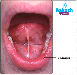
Fig: Frenulum
Lingual papillae
Papillae are projections present on the upper surface of the tongue. Some of them bear taste buds. They are classified mainly into four types as follows:
Circum -vallate papillae
They are the largest lingual papillae. They are arranged normally in an inverted V-shaped row. It is present in the base of the tongue. They are 8-12 in number. Each of this papillae is surrounded by a circular groove and contains around 100 taste buds. They possess gustatory receptors.
Filiform papillae
They are the smallest papillae. They are numerous and occur mainly near the centre. They possess tactile or touch receptors.
Fungi-form papillae
They are rounded papillae. They are numerous. They are present near the tip. They possess around five taste buds.
Foliate papillae
They are not developed in humans. They are present in other mammals mainly on the sides of the tongue.
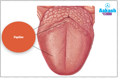
Fig: Papillae
Taste buds
They are considered as the receptors of taste that are present within some papillae. An average person has about 10,000 taste buds.
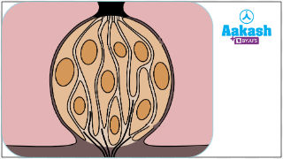
Fig: Taste buds
Tastes
They sense all four tastes like bitter, sour, salty, and sweet and send messages to the brain to generate the sensation of taste. Umami taste is considered as the fifth taste. It means the essence of deliciousness in Japanese and it tastes meaty or savoury.
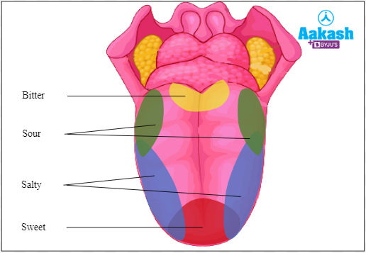
Fig: Tastes
Functions of the tongue
- The food is lodged between the grinding surfaces of teeth with the help of teeth.
- It helps in swallowing the food.
- It helps in removing the waste materials from the teeth.
- It aids in speech along with lips, teeth and palate.
Teeth
Teeth are hard structures that are present in two semicircles. They are embedded in the socket of the jaw bone. Teeth are eco-mesodermal in origin. They are of several uses for teeth like gripping, gnawing, cutting, tearing and crushing.
Fig: Teeth
Parts of tooth
The tooth is composed of the following parts:
Crown
It is the exposed part of the tooth. It projects above the gums.
Enamel
It is the hardest part of the human body that covers the crown. Enamel is secreted by the ameloblast cells. It is acellular, avascular and non-regenerable. The function of enamel is to masticate the food.
Neck
It is the middle part of the tooth which is surrounded by the gums.
Root
It is considered as the basal part of the tooth. It is seen embedded in the jaw bone.
Dentine
The crown and root of the teeth are made up of dentine.
Pulp cavity
It is the cavity enclosed by the dentine. It possesses a gelatinous, soft, connective tissue called pulp. It possesses specialised dentine forming cells which are called odontoblasts.
Pulp canal
It is the canal through which blood supplies and nerves enter the pulp cavity.
Periodontal membrane
It helps in fixing the root to the jaw bone.
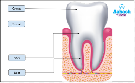
Fig: Parts of the tooth
Classifications of animal teeth
There are three classifications of animal teeth based on structure and function, placement in jaw and appearance in life.
Classification of dentition on the basis of structure and function
On the basis of structure and function, teeth are classified into two types as follows:
Homodont teeth
A single type of tooth is present in the oral cavity in this condition. Examples include the teeth of dolphins.
Heterodont teeth
In this particular condition different types of teeth are seen present in the oral cavity. Examples include human beings and cheetahs.
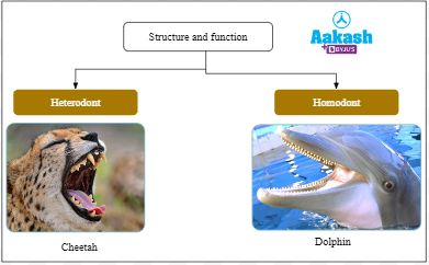
Fig: Types of teeth on the basis of structure and function
Types of human teeth
They are of four types as follows:
Incisors
They are considered chisel shaped and possess one root. They are eight in number and mainly used for cutting.
Canines
They are dagger-shaped and have one deep root. They are four in number and used for ripping and shredding.
Premolars
They are the bicuspid teeth because they have two cusps at the crown. The premolars of the upper jaw possess two roots while the premolars of the lower jaw have only one root. They are normally eight in number and used for crushing and chewing.
Molars
On the upper jaw they possess three roots while the molars of the lower jaw possess two roots. They are 12 in number. They help in crushing, chewing and grinding.
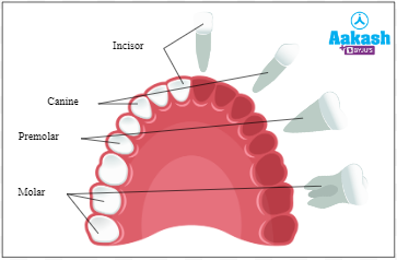
Fig: Types of human teeth
Classification of dentition based on their placement in the jaw
On the basis of placement in the jaw, they are of three types as follows:
Thecodont dentition
These types of teeth are normally present in the bony sockets of the jaw bone. They are found in man, crocodile etc.
Pleurodont dentition
These types of teeth are normally present on the lateral side of the jaw bone. They are most common in reptiles.
Acrodont dentition
These types of teeth are present normally on the terminal part of the jaw bone. These are found in amphibians and fish.
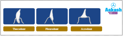
Fig: Types of teeth on the basis of placement in jaw
Classification of dentition on the basis of appearance in life
On the basis of appearance in life, teeth are of three types as follows:
Monophyodont
These types of teeth appear once in lifetime. Examples include wisdom teeth, that is the third molar in man which appears once in lifetime.
Diphyodont
Humans possess two sets of teeth, milk teeth and permanent teeth. This condition is called diphyodont dentition.
Polyphyodont
These types of teeth appear more than twice in the lifetime of an organism. Examples include teeth in amphibians.
Classification of human dentition based on the appearance in life
On the basis of their appearance in life, human teeth are of two types as follows:
Temporary teeth
They are also known as deciduous or milk teeth. They are developed fully by the age of two or three years in human beings. They are 20 in number. Premolars are absent in temporary teeth. Dental formula of temporary teeth is shown below:
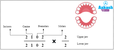
Fig: Dental formula of temporary teeth
Permanent teeth
They replace the temporary teeth. They start forming from the age of six and complete formation in normal instances by the age of 12. The last molar or wisdom teeth form usually after the age of 18. They are 32 in number. Once broken, they cannot be replaced naturally. Dental formula of permanent teeth is shown below:
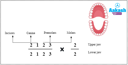
Fig: Dental formula of permanent teeth
Pharynx
Pharynx is a tube of 12-14 cm in length. It is situated behind the soft palate. Pharynx provides a common passage for food and air. It helps in swallowing the food. The muscles of the pharynx contract. This raises and expands the lumen of pharynx that helps in swallowing. It is divided into three parts as follows:
- Nasopharynx
- Oropharynx
- Laryngopharynx
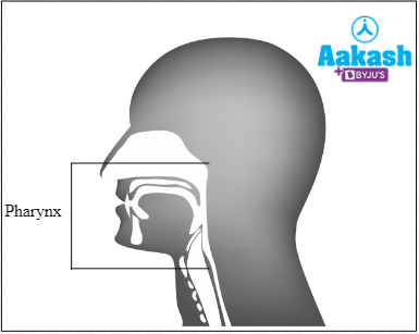
Fig: Location of pharynx
Nasopharynx
It lies behind the nasal chambers above the soft palate. It is connected with the middle ear through the eustachian tube. This part has tonsils called pharyngeal tonsils or adenoids. Tonsils provide the first line of defence against illness. White blood cells are produced there that help in fighting the infections.
Oropharynx
It is the middle part of the pharynx. It lies behind the oral cavity. The palatine tonsil, an ovoid mass of lymphoid tissue is present here. Tonsillectomy is done here to remove the infected tonsil tissue.
Laryngopharynx
It leads to the oesophagus and possesses glottis and epiglottis. The wall of pharynx has voluntary constrictor muscles which force the food in the form of bolus into the oesophagus. The lymphatic tissue of the pharynx arranged in the form of a ring called Waldeyer’s ring.
Epiglottis
Epiglottis is a cartilaginous flap located in the throat. The function of epiglottis is to prevent the food from entering the glottis (opening of the windpipe) while swallowing.
GIF: Movement of food in the pharynx
Oesophagus
It has a tube-like structure that is narrow and muscular. It extends from the posterior side of the neck, thorax and diaphragm. It is about 25 cm long and two cm wide. The other names of oesophagus are food pipe, gullet or the food tube. It is divided into three parts as follows:
Cervical part
It is present in the neck region.
Thoracic part
It is present in the thorax region.
Abdominal part
It is present in the abdominal region.
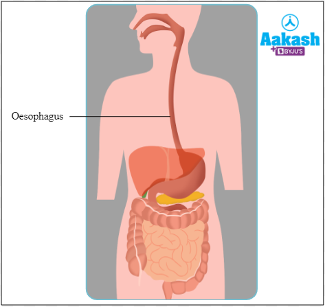
Fig: Location of oesophagus
It transfers food from the pharynx to the stomach through a specialised movement called peristalsis. Peristalsis is a series of wave-like muscle contractions that move food. Muscles in the esophagus prevent entry of air into the digestive tract.
GIF: Peristalsis
Stomach
The stomach is a J-shaped organ in the upper-left corner of the abdomen. It is a hollow organ that retains food for up to 4 - 5 hours before digesting it. Its inner surface is highly convoluted, allowing it to fold up when empty and expand like a balloon when the stomach is filled with food.
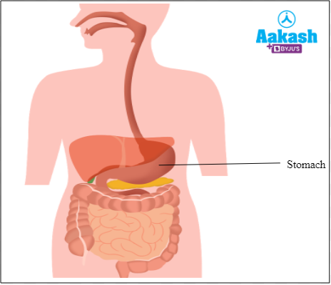
Fig: Stomach
Parts of stomach
Stomach is divided into four parts as follows:
- Cardiac
- Fundus
- Body
- Pylorus
Cardiac
In the cardiac region, the oesophagus connects to the stomach. The opening of the oesophagus to the stomach is called cardiac aperture. The cardiac region refers to the portion of the stomach that is located near the heart.
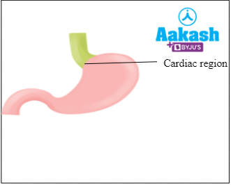
Fig: Cardiac part of stomach
A gastro-oesophageal sphincter or cardiac sphincter guards the entry of the oesophagus into the stomach, preventing food from returning to the oesophagus. A sphincter is a specialised muscle that opens or closes an entrance or passage in the body by relaxing. This sphincter is less effective in infants.
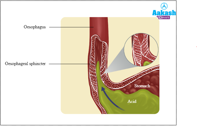
Fig: Gastro-oesophageal sphincter
Fundus
This region is filled with air and gas. It is a small dome shaped part.
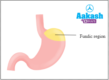
Fig: Fundic part of the stomach
Body
This is the middle and main part of the stomach. Food is broken down into smaller particles because the enzymes act here.
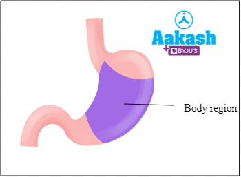
Fig: Body part of stomach
Pylorus
Pylorus is also known as the antrum. It is the posterior or lowest part of the stomach that opens into the duodenum part of the small intestine through the pyloric aperture. This opening is guarded by the pyloric sphincter. It possesses a wider pyloric antrum and a narrow pyloric canal.
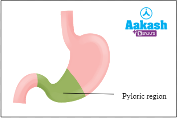
Fig: Pyloric part of stomach
Functions of the stomach
- It helps in the storage of food.
- It helps in the churning and breaking down of food.
- It regulates the flow of food into the small intestine.
Small Intestine
Small intestine is a tube-like structure which is long, narrow and coiled. In adults it is about 6.25 metres long. It is divided into three parts, duodenum, jejunum, and ileum.
Duodenum
Duodenum is the shortest and widest part of the small intestine. It is a C-shaped structure. It receives the hepatopancreatic ampulla or Ampulla of vater.
Jejunum
It is considered as the middle part of the small intestine. It is a coiled tube with a diameter of about 4 cm and 2.5 cm long. It is a muscular part and therefore, it is reddish in appearance.
Ileum
Ileum is the longest part of the small intestine. It is a highly coiled structure with a diameter of 3.5 cm and 3.5 m long. The wall of the ileum is thinner as compared to the wall of the jejunum.
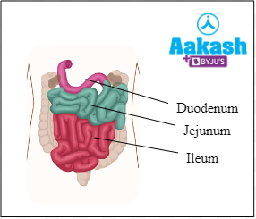
Fig: Small Intestine
Peyer’s patches
Peyer patches are a unique feature of the ileum that are named after Johann Conrad Peyer. These are characterised by clusters of the lymphatic tissue. The lymphatic tissue produces lymphocytes.
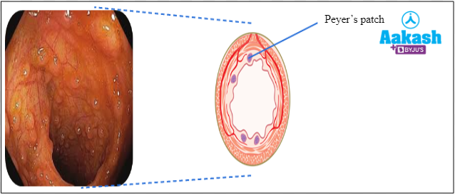
Fig: Peyer’s patches
Plicae circulares
These are the prominent structures in jejunum. Plicae circulares are characterised as circular folds and also known as valves of Kerckring. They increase the surface area for efficient absorption.
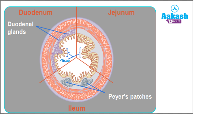
Fig: Structures in the small intestine
Villi
Intestinal villi are the finger-like projections that increase the surface area for absorption.
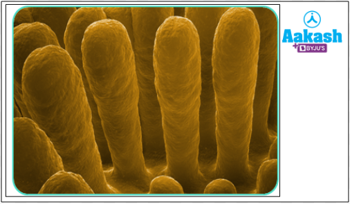
Fig: Villi in small intestine
Intestinal villi is made up of mucosa layers. It increases surface area for absorption. It is covered by epithelium. Villi have lacteal (lymph vessel) and blood capillaries.
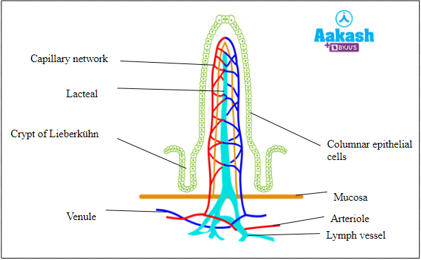
Fig: Structure of villi
Microvilli
Microvilli are microscopic projections present in a single villus. They collectively give a brush border appearance. They increase the surface area for absorption of food. These structures are absent on Peyer’s patches. Blood vessels and lacteals are situated close to the microvilli. The nutrients from the microvilli are transported to the blood vessels and lacteals.
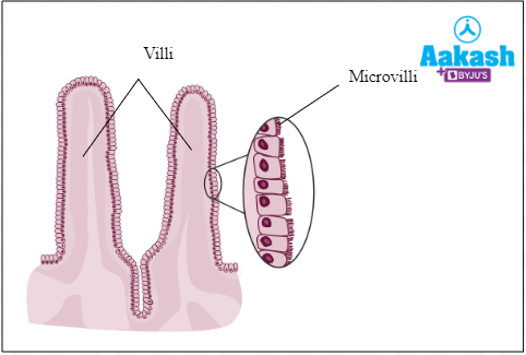
Fig: Structure of microvilli
Functions of small intestine
- In this region the digestion of food is completed.
- It helps in the absorption of food into the blood and lymph.
- It secretes some hormones like cholecystokinin, secretin etc.
Large Intestine
The large intestine is known because of its large diameter. It is a tubular structure which is about 1.5 m long. The large intestine is divided into three parts: caecum, colon and rectum.
Caecum
Caecum has a characteristic pouch-like structure into which ileum opens. It is host to symbiotic microbes. There is a large blind sac that opens into the colon. It is 6cm wide. At the ileocaecal junction a valve called ileocolic valve is present. It is well- developed in herbivores and helps in the digestion of cellulose by bacterial action. It regulates the passage of materials from the small to the large intestine. Caecum has a vermiform appendix.
Vermiform appendix
It is a narrow finger-like projection. It is about 8cm long. It is considered as a vestigial organ. It is a residual part from ancestors with no known function. Inflammation of the appendix is known as appendicitis.
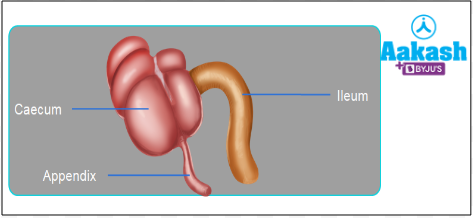
Fig: Caecum
Colon
Caecum leads to colon. Colon is responsible for the reabsorption of fluids. It processes waste products and prepares for elimination from the body. Colon is composed of four parts as follows:
- Ascending colon - It passes upwards.
- Transverse colon - It runs across the abdominal cavity.
- Descending colon - It descends downwards.
- Sigmoid colon - It joins with the rectum.
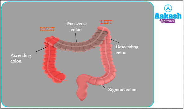
Fig: Colon
Rectum
Rectum is considered the last part of the digestive tract. It ends in anal canal that opens through anus. It is about 15 - 20 cm long. Piles or haemorrhoids normally develop in this region. The bowel movement of anus is controlled by two sphincters:
- Internal anal sphincter has involuntary and smooth muscle fibres.
- External anal sphincter has voluntary and striped muscle fibres.
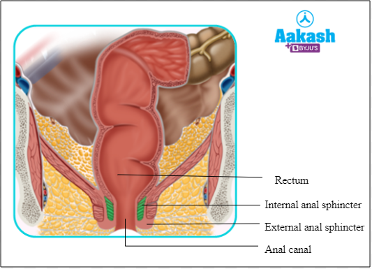
Fig: Rectum
Functions of large intestine
- It temporarily stores food.
- It helps in the elimination of faeces.
- It helps in the absorption of vitamin K and vitamin B complex.
Histology of digestive system
The alimentary canal is composed of four basic layers as follows:
- Serosa
- Muscularis
- Submucosa
- Mucosa
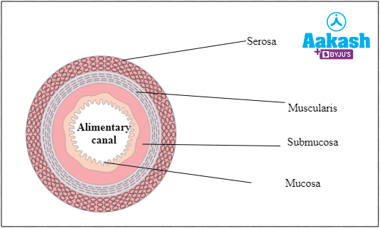
Fig: Layers of alimentary canal
Serosa
It is the outermost layer of the stomach and is also known as visceral peritoneum. It is composed of thin squamous epithelium also called mesothelium along with some connective tissue.
Muscularis externa
This layer is formed by smooth muscles. The cells of the muscularis layer are arranged into inner circular and outer longitudinal layers. In the stomach region an additional layer of oblique muscle fibres is also present. To control the muscular contractions, a network of nerve cells called Auerbach’s plexus or myenteric plexus is present between the longitudinal and circular muscle fibres.
Submucosa
It is formed of loose connective tissues. It possesses nerves, lymph vessels and blood vessels. The intestinal glands are present in this region. A network of nerve cells called Meissner’s plexus or submucosal plexus lies between the muscular coat and the submucosa.
Mucosa
It is the innermost lining of the lumen of the alimentary canal. It contains secretory and absorptive cells. In the stomach, it forms irregular folds known as rugae. This layer has mucus-secreting goblet cells. Mucus helps in lubrication. Rugae increase the surface of the stomach and disappear when the stomach distends.
Practice Problems
Q1. Match Column A with Column B.
|
Column A |
Column B |
|
P. Colon |
|
Q. Cardiac |
|
R. Ileum |
A. 1 - R, 2 - Q, 3 - P
B. 1 - Q, 2 - R, 3 - P
C. 1 - Q, 2 - P, 3 - R
D. 1 - P, 2 - R, 3 - Q
Solution: The stomach is a J-shaped organ in the upper-left corner of the abdomen. In the cardiac region, the oesophagus connects to the stomach. The cardiac region refers to the portion of the stomach that is located near the heart.
Small intestine is a tube-like structure which is long, narrow and coiled. Ileum is considered as the longest part of the small intestine. It is a highly coiled structure with a diameter of 3.5 cm and 3.5 m long.
Large intestine is known because of its large diameter. It is a tubular structure which is about 1.5 m long. Colon is responsible for the reabsorption of fluids. It processes waste products and prepares for elimination from the body. Hence, the correct option is b.
Q2. Which of the following structures serve in the respiratory system as well as digestive system?
A. Pharynx
B. Tongue
C. Rectum
D. Villi
Solution: The pharynx is an important element of the digestive and respiratory systems. It is a hollow structure lined with a moist tissue. It takes in air from the nasal tube and food and air from the mouth. The pharynx transports food and air to the oesophagus and larynx, respectively. Hence, the correct option is a.
Q3.. Match the following:
|
Layers of alimentary canal |
Description |
|
i. Innermost layer contains irregular folds called rugae |
|
ii. Composed of loose connective tissue and contains lymph, blood and nerves |
|
iii. Outermost layer and composed of mesothelium and connective tissue |
|
iv. Composed of smooth muscles and longitudinal from outside and circular from inside |
A. 1 - i, 2 - iii, 3 - iv, 4 - ii
B. 1 - ii, 2 - ii, 3 - iv, 4 - iii
C. 1 - iv, 2 - i, 3 - iii, 4 - ii
D. 1 - iii, 2 - iv, 3 - ii, 4 - i
Solution: the alimentary canal is composed of four layers. These are serosa, muscularis externa, submucosa and mucosa.
The outermost layer is the serosa. It is made up of connective tissue and mesothelium.
Smooth muscles form the muscularis layer. It has a longitudinal muscle layer on the outside and a circular muscle layer on the inside.
The submucosa is made up of connective tissues that are loose. It is made up of lymph, blood, and nerves.
The mucosa is the layer closest to the skin. In the stomach region, it has uneven folds and gastric glands. Hence, the correct option is d.
Q4. Name the parts of the large intestine.
Answer: The large intestine is classified into three parts. These are as follows:
- Caecum
- Colon
- Rectum
Q5. What are the different parts of the colon?
Answer: Colon is composed of four parts as follows:
- Ascending colon
- Transverse colon
- Descending colon
- Sigmoid colon
Q6. Explain the structure of microvilli?
Answer: Microvilli are microscopic projections present in a single villus. They collectively give a brush border appearance. They increase the surface area for absorption of food. These structures are absent on Peyer’s patches. Blood vessels and lacteals are situated close to the microvilli.
FAQs
Q1. Where is protein first digested?
Answer: Protein digestion starts in the stomach. Here the pepsin acts on proteins and converts them into smaller peptides.
Q2. Why are humans unable to digest cellulose?
Answer: Humans cannot digest cellulose because they lack enzyme cellulase that is required for breaking the beta-acetal linkages of cellulose.
Q3. What is the size of an empty stomach?
Answer: The human stomach is about the size of a fist when there is no food in it.
Q4. What causes hiccups?
Answer: Hiccups are caused by spasms in the diaphragm, a small muscle that helps to breathe. When this happens, the air is quickly drawn down into the stomach, and we burp it out again. Hiccups can occur frequently and are triggered by a variety of factors, including consuming too much food.