-
Call Now
1800-102-2727


Cell wall, Practice Problems and FAQs
Can you imagine a home without walls? In fact no building is complete without walls. Walls not only help to maintain the structure of a building they also protect the inhabitants of a building from the dangers of the world. The stronger the protection needed, the stronger and bigger the wall will be. Right? Thus, we can conclude that the main functions of a wall are protection and maintaining a structure.
You will be amazed to know that many living beings have a similar wall protecting the contents of the cells in their body. It is the cell wall. The function of this wall is pretty much similar to the walls of the buildings but structurally it is very different. In fact the composition and structure of the cell wall varies from one group of organisms to another. The presence of a cell wall provides firmness and rigidity to the cells so that they can be protected from the external forces. So, why don't animals have a cell wall? This is because in order to carry out movement and locomotion from one place to another, animals need a higher degree of flexibility in their body and hence their cells. If the cell wall would have been present in animal cells, their bodies would also have been stiff like plants.

In this article we will discuss the structure and function of cell walls present in different types of organisms and understand how different are prokaryotic cell walls from the eukaryotic ones.
Table of contents:
- Cell wall
- Prokaryotic cell wall
- Eukaryotic cell wall
- Functions of cell wall
- Practice Problems
- FAQs
Cell wall
The thick porous outer protective covering in bacteria, fungi and plants is known as cell wall. It can provide rigidity and strength to the cells. Another major function of the cell wall is to facilitate cell to cell interaction. Cell walls are absent in animal cells. The structure of the cell wall is different in prokaryotes and eukaryotes.
Prokaryotic cell wall
The prokaryotes are those organisms which are made up of cells that lack a well-defined membrane bound nucleus and other membrane bound organelles. All the prokaryotic organisms are unicellular. The prokaryotic cells are the most primitive cells which appeared about 3.5 billion years ago. Since they are the most primitive ones, the cell wall is also not advanced like eukaryotes. The outer covering of a prokaryote is called a cell envelope. The cell wall of a prokaryote is a part of the cell envelope.
Fig: Parts of cell envelope

Structure of prokaryotic cell wall
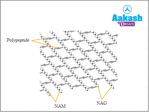
The cell walls of prokaryotes give them proper shape and support. It is made up of a polymer called peptidoglycan which contains polysaccharides and amino acids. It has a highly cross-linked structure which provides strength to the cell wall. Peptidoglycan is a polymer of alternating residues of N- acetylglucosamine (NAG) and N- acetyl muramic acid (NAM). A small peptide side chain or 3-5 amino acids is attached to NAM and it cross links with the peptide chain of another strand, thus forming a mesh like structure in three dimensions.
Fig: Structure of peptidoglycan
In some bacteria teichoic acid is also a component of the cell wall. Mycoplasma is the only prokaryote that lacks a cell wall. The composition of the cell wall varies in gram negative and gram positive bacteria.
Fig: Cell wall of bacteria

Gram positive and Gram negative bacteria
Bacteria is divided into Gram positive and Gram negative based on their reaction to Gram staining. Hans Christian Gram invented this method. This difference in the response to Gram staining is due to the difference in their cell wall structure. Gram positive bacteria do not have an outer membrane of lipoproteins and lipopolysaccharides as do Gram negative bacteria.
Fig: Gram negative and gram positive bacteria

Gram staining is a process of adding crystal violet to the bacterial cell culture mounted on to the slide. It is followed by addition of Gram’s iodine, to fix the colour and then washed off with alcohol after a minute. At the end safranin is added to the mount as a secondary stain. After this process, the slide is observed under the microscope. Bacterial cells which retain the colour of the primary stain or crystal violet and appear blue are said to be Gram positive. The ones which take up the colour of the secondary stain, that is safranin, and appear red are said to be Gram negative.
Fig: Gram staining

Cell wall of Gram positive bacteria
The cell wall of a Gram positive bacteria is single layered and is mostly composed of a thick layer of peptidoglycan. It has a thickness of 20 nm to 80 nm. The thick layer of highly cross linked peptidoglycan is responsible for trapping the primary stain, that is, crystal violet. Hence, it is not lost even after washing with alcohol and is thereby not replaced by the pink colour of safranin which is applied later.
The rest of the cell wall is composed of teichoic acids. These teichoic acids are linked with lipids in the plasma membrane through covalent bonds to form lipoteichoic acids. They help in the anchoring of the cell wall to the cell membrane. They have no porins on the cell wall. Some examples of gram positive bacteria are, Actinomyces, Clostridium, Mycobacterium, Streptococci, Staphylococci etc.
Fig: Cell wall of Gram positive bacteria

Cell wall of Gram negative bacteria
The cell wall of Gram negative bacteria has a thinner layer of peptidoglycan and is surrounded by a layer of lipoproteins and lipopolysaccharides. During the alcohol wash, the lipid rich layer dissolves in the alcohol and hence the primary stain or crystal violet is also washed off. Thus these bacterial cells pick up the pink colour after staining with safranin.
Teichoic acid is absent in the cell wall of Gram negative bacteria but porins are present. Some examples of Gram negative bacteria are Salmonella, Pseudomonas, Vibrio, Rhizobium, Escherichia etc.
Fig: Cell wall of Gram negative bacteria

Eukaryotic cell wall
Except the animals, most of the eukaryotic organisms have cell walls in their cells. The function of the cell wall is protection and rigidity, but the composition of the cell wall varies in different eukaryotes. It depends on their mode of nutrition, ability to move, habitat etc. About 2.7 billion years ago eukaryotic cells evolved from prokaryotic cells. So there will be some similarities and also differences between prokaryotic and eukaryotic cells and so their cell wall.
Fig: Plant cell

Structure
The cell wall has a highly intricate lattice structure. It possesses three types of networks - cellulose microfibrils, pectic polysaccharides and structural proteins. The protein network interweaves through the other two networks. The cell wall consists of three main parts - matrix, microfibrils and cell wall depositions.
Matrix
The matrix is a highly crosslinked amorphous gel-like substance composed of water (30%-60%), pectin (2%-8%), hemicellulose (5%-15%), lipids (0.5%-3%) and proteins (1%-2%).
Pectin forms the colloidal complex and filler substance of the matrix and cross-links different substances. It determines the hydration of the wall, its elasticity, microfibril orientation and growth.
Hemicellulose is a heteropolysaccharide that binds the microfibrils to the matrix of the cell wall. Lipids provide linkage to the hydrophobic ends of the cell wall depositions and also help in the transport of hydrophobic substances across the wall. Proteins present in the cell wall are mostly structural proteins except for some which are enzymes.
Microfibrils
These are the structural elements of the cell wall and provide strength and rigidity to the cell wall much like steel bars and mesh used in concrete ceilings. In plants the microfibrils are formed of cellulose. Each microfibril consists of 2000-2500 parallel straight cellulose chains. Depending upon the type of the plant cell, cellulose microfibrils may form 20-40% of the cell wall. Microfibrils are loose and wavy in the primary cell wall of plants while they have a close and parallel arrangement in the secondary cell wall. Microfibrils in fungal cell walls are composed of chitin.
Cell wall depositions
These are the addition of organic and inorganic substances into and on the outer side of the cell wall. Some of the commonly deposited materials are silica, non-siliceous minerals such as iron, calcium, etc., cutin (found in cuticle of leaves and stems), suberin (found in cork cells), waxes (also found in cuticle) and lignin (found in sclerenchymatous cells).
Cell wall of fungi
The fungal cell wall has a dynamic structure which protects the fungi from environmental stress. It also helps to live in varying osmotic pressure. The process of production of complex molecules, i.e, biosynthesis is an important feature of the fungal cell wall. Hence they have a complicated cell wall structure. Most pigments are present in the cell wall and protects the fungus from the harmful ultraviolet rays. The major composition of fungal cell walls are as follows:
- Glucans
- Chitin
Fig: Fungal cell

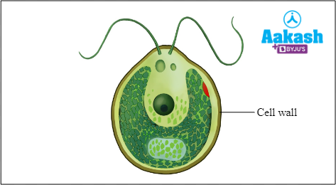
Cell wall of algae
The cell wall of algae is composed of fibrils, matrix and crystalline polymers that can interact with water. The cell walls of different algae are of different types. The common cell wall composition of algae are as follows:
- Cellulose
- Galactans
- Mannans
- Minerals like calcium carbonate
Fig: Algal cell
Apart from this the cell wall also has the extracellular matrix as the dominant component and the carbon fixed through photosynthesis. Let’s see how different the cell walls of different algae are:
- Multi shaped scales are seen in the cell walls of primitive algae of Prasinophyceae.
- The cell walls of charophycean green algae contain polymers like land plants such as cellulose, hemicellulose, etc..
- The red algae has a cell wall made up of cellulose, xylan or mannan fibrils and extensive matrix polysaccharides.
- Silica based cell walls are seen in diatoms which are unicellular algae placed in kingdom Protista.
- The cell wall of brown algae contains cellulose, matrix polysaccharides and phenolics.
Cell wall of plants
The outermost covering of the plant cell is called a cell wall. Since higher plants are more advanced than algae and fungi, the structure of their cell wall is also advanced. The cell wall is mainly composed of some insoluble carbohydrates like cellulose, hemicellulose, pectin and proteins. The percentage of the cell wall components and its thickness highly varies according to the type of plant or cell. In a young plant cell, there will be only a primary cell wall. When the pants grow, the secondary walls are formed inside the primary cell wall.
Layers of the plant cell wall
The physical structure of plant cell wall is composed of three main layers as follows:
- Middle lamella
- Primary cell wall
- Secondary cell wall
The adjacent cells of a plant are connected through the protoplasmic connections, known as plasmodesmata, that run across pores present in the cell wall. These structures are also important when we discuss the structure of a plant cell wall. Let us now discuss how these regions are supporting the cell wall of the plant.
Fig: Structure of plant cell wall
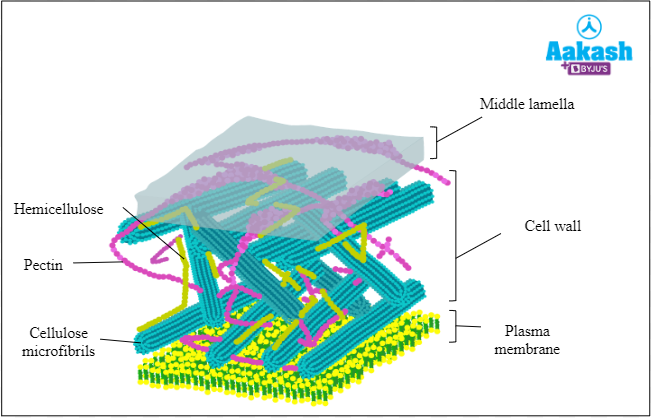
Middle lamella
The amorphous structure of a plant tissue which can glue together the adjacent cells is known as middle lamella. It is made up of calcium and magnesium pectate (simply called pectins). During cytokinesis (division of cytoplasm during cell division), the middle lamella is the first formed layer. It is a common layer to all cells.
Fig: Middle lamella

Primary cell wall
The first formed wall of the cell is called the primary cell wall. It is usually seen in young plants. It is thin and capable of extension. It grows by intussusception (new cell wall components are added within the existing components).
Secondary cell wall
The cell wall formed after the primary cell wall and inner to the primary cell wall is called a secondary cell wall. It is found in mature cells. It is thick and usually made up of three or more layers. New wall components are layered on to the inner surface of the wall.
There are three layers in the secondary wall of plants. They are S1, S2 and S3. The major difference between these layers is that the direction of the arrangement of cellulose microfibrils are different.
Fig: Layers of secondary cell wall

Difference between primary and secondary plant cell wall
Primary cell wall |
Secondary cell wall |
|
Hemicellulose: 50% |
Hemicellulose: 25% |
|
Lipids: 5-10% and Proteins: 5% |
Lipids and proteins in very low quantity or absent |
|
Formed by Intussusception |
Formed by Intussusception and Apposition |
|
Universally found in all plant cells |
Absent in meristematic cells |
Pits
Pits are unthickened areas or depressions in the secondary walls of plant cells. Pits of the adjacent cells generally occur exactly opposite and form pit pairs. A pit present on the free surface of the cell will be without its corresponding partner and is known as a blind pit.
A pit is made up of a pit chamber and a pit aperture. The pit chamber or pit cavity is the depression in the wall, representing the area where the secondary wall is absent. Opening of the pit chamber into the cell is known as the pit aperture. Pit membrane is a permeable membrane which forms the floor of the pit and is formed of the primary wall and middle lamella between adjacent cells. Minute submicroscopic pores may occur in the pits.
Pits can be simple or bordered. Simple pits have pit chambers with uniform width similar to that of the pit aperture. In bordered pits, the pit chamber is flask shaped and narrows towards the pit aperture. The secondary cell wall therefore overarches the pit chamber.
Fig: Bordered pits

Plasmodesmata
The cytoplasmic bridges between the adjacent plant cells is known as plasmodesmata. They develop in the minute pores that run across the middle lamella and the cell walls. They help to establish connections between the cytoplasm of the adjacent cells.
Fig: Plasmodesmata

Functions of cell wall
The cell wall performs many functions. Some of them are as follows:
- Maintains the size and shape of the cell.
- Protects the cell from shocks and mechanical injuries.
- Helps to protect the cell from pathogen attacks.
- Being freely permeable, it allows ransfer of materials in and out of the cell.
- Cutin and suberin deposition in the cell wall helps reduce the loss of water through transpiration.
- Plasmodesmata ,the cytoplasmic extensions of plant cells, link up all the protoplasts of a tissue.
- Growth of the cell wall helps the cell to enlarge its size.
Practice Problems
Q 1. Assertion: The gram positive bacteria appears blue when observed under the microscope after Gram staining.
Reason: Gram positive bacteria have thin cell walls.
Which of the following statements is correct about the assertion and reason given above?
a. Both assertion and reason are true and reason is the correct explanation of assertion.
b. Both assertion and reason are true but reason is not the correct explanation of assertion.
c. The assertion is true, but the reason is false.
d. Both assertion and reason are false.
Answer: Bacteria is divided into gram positive and gram negative based on their reaction to gram staining. Hans Christian Gram invented this method. This difference in the response to gram staining is due to the difference in their cell wall structure. The cell wall of a Gram positive bacteria is single layered and is mostly composed of a thick layer of peptidoglycan. It has a thickness of 20 nm to 80 nm. The thick layer of highly cross linked peptidoglycan is responsible for trapping the primary stain, that is, crystal violet. Hence, it is not lost even after washing with alcohol and is thereby not replaced by the pink colour of safranin which is applied later. Hence the correct option is c.
Q 2. Match the following organisms with their cell wall composition.
|
I) NAM and NAG |
|
II) Cellulose, hemicellulose and pectin |
|
III) Galactans and mannans |
|
IV) Glucans and chitins |
a. A-I, B-II, C-III, D-IV
b. A-I, B-II, C-IV, D-III
c. A-II, B-IV, C-III, D-I
d. A-II, B-I, C-IV, D-III
Answer: The outermost covering of the plant cell is called a cell wall. The cell wall is mainly composed of some insoluble carbohydrates like cellulose, hemicellulose, pectin and proteins. The percentage of the cell wall components highly varies according to the type of plant or cell.
The cell walls of prokaryotes give them proper shape and support. The cell wall is made up of peptidoglycans which contain polysaccharides and amino acids. Peptidoglycan is a polymer of alternating residues of N- acetylglucosamine (NAG) and N- acetyl muramic acid (NAM). A small peptide side chain or 3-5 amino acids is attached to NAM and it cross links with the peptide chain of another strand, thus forming a mesh like structure in three dimensions.
The fungal cell wall has a dynamic structure which protects the fungi from environmental stress. The major composition of fungal cell walls are glucans and chitins.
The cell wall of algae is composed of fibrils, matrix and crystalline polymers that can interact with water. The cell walls of different algae are of different types. The common cell wall composition of algae are cellulose, galactans, mannans and minerals like calcium carbonate. Hence the correct option is d.
Q 3. Which of the following statements are incorrect about the functions of a cell wall?
I) Maintains the shape and size of the cell
II) Helps to keep the cell from pathogen attacks
III)Plasmodesmata link up all the protoplasts of a tissue
IV) Cutin and suberin deposition in the cell wall helps to enlarge the size of the cel
a. I
b. II
c. III
d. IV
Answer: The cell wall performs many functions. It maintains the shape and size of the cell. It can protect the cell from shocks and mechanical injuries and also helps to keep the cell from pathogen attacks. Cutin and suberin deposition in the cell wall helps reduce the loss of water through transpiration. Plasmodesmata, the cytoplasmic connections between adjacent plant cells, link up all the protoplasts of a tissue. Growth of the cell wall helps the cell to enlarge its size. So here the incorrect statement is IV. Hence the correct option is d.
Q 4. What is the middle lamella of a plant cell wall?
Answer: Middle lamella is one of the physical structures of plant cells. It is the amorphous structure of a plant tissue which can glue together the adjacent cells. It is made up of calcium and magnesium pectate (simply called pectins). During cytokinesis (division of cytoplasm during cell division), the middle lamella is the first formed layer. It is a common layer to all plant cells.
FAQs
Q 1. Cell wall of a plant is living or dead?
Answer: Cellulose is the major component of the plant cell wall. It is a polymeric carbohydrate which provides rigidity to the cell walls. Since cellulose is a non living component the cell wall is also non living. The cell wall does not show any internal activity and it has the function of protecting the internal protoplasm of the cell.
Q 2. Can a cell survive without a cell wall?
Answer: There are some bacteria which can retain their ability to live inside living organisms, even after they lose the cell wall. They have tough cytoplasmic membranes which can resist external pressures. Hence we can say that bacterial cells can survive without a cell wall. There are some bacteria like Mycoplasma and Ureaplasma which lack cell walls.
Q 3. Do viruses have a cell wall?
Answer: No, viruses do not have a cell wall. They have a protective protein coat instead of a cell wall. This protein coat covers the nucleic acid inside it and the coat is called capsid.
Q 4. What are the economically important cell walls?
Answer: The fibres like cotton and linen are obtained from the walls of long and strong fibre cells of the plants. Other materials such as lumber, charcoal and some wood products are also the economically important products from the cell wall. Carrageenan and agar are two products from algal cell walls. These are included in the extensive matrix polysaccharides of the cell wall of red algae.
YOUTUBE LINK:https://www.youtube.com/watch?v=-NOY_k8iN9A
Related Topics
|
What is a cell?: Discovery of cell, Characteristics of typical cell, Practice Problems and FAQs |
|
Prokaryotic Cell: Overview, Practice Problems, FAQs |
|
Eukaryotic Cell, Practice problems and FAQs |