-
Call Now
1800-102-2727


Differences Between Chromosomes and Chromatids, Practice Problems and FAQs
We all know that all the activities in our body are under the control of the brain. It is considered as the central processing unit of our body. Our body is made up of cells which are the structural and functional unit. Now can you tell me which organelle controls all the activities of the cell? Yes, you are correct, it is the nucleus. The nucleus possesses all the information required for controlling metabolism and transmission of characters from one generation to the next generation. But where all this information is exactly present in a cell? Yes these informations are present in the genetic material which exists in the form of chromosomes. Now you have lots of queries related to chromosomes in your mind. In this article we will see the main differences between the chromosomes and chromatids.
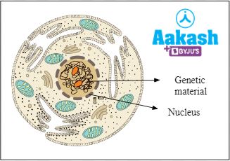
Fig: Cell with nucleus
Table of contents
- Chromosomes
- Chromatids
- Differences between chromosomes and chromatids
- Practice Problems
- FAQs
Chromosomes
Chromosomes are the thread-like structures present inside the nucleus of eukaryotic cells like, fungal, plant and animal cells. The chromosomes are believed to be discovered first by Hofmeister. But the term ‘chromosome’ was coined by Waldeyer in 1888.
Composition of chromosomes
These are composed mainly of proteins and DNA. They appear threadlike or rod-shaped inside the nucleus. These are the condensed forms of chromatin fibres and can be stained. These act as hereditary vehicles by storing and transmitting hereditary information from one generation to the next generation. Chromosomes appear under a light microscope during the karyokinesis (division of nucleus) phase of cell division. The number of chromosomes are fixed in a species. These are present in single sets (n) in haploid forms or gametophytes. But are present in two sets in diploid forms or sporophytes. For example, eggs possess 23 chromosomes (n) and somatic cells possess 46 chromosomes (2n) in human beings.
History of chromosome
The word chromosome means ‘colourful bodies’. These are visible under a microscope once stained. ‘Chroma’ means colour and ‘soma’ means body. Walther Flemming, in 1879, used basic stains to study the cell division in cells of Salamander. He observed some intensely stained parts in the nucleus and called them ‘chromatin’.
Fig: Walther Flemming
Thomas Hunt Morgan identified the relationship between the X chromosome with gender and eye colour in fruit flies or Drosophila melanogaster in the 1900s.
Fig: Thomas Hunt Morgan
Histone proteins and non histone proteins bind with the large DNA molecules and help in condensation to form chromosomes. This helps in the proper packaging of long DNA molecules inside the nucleus of a cell.
Structure of chromosome
A typical chromosome consists of the following parts:
- Chromatids
- Centromere
- Kinetochore
- Telomere
- Secondary constriction
- Matrix
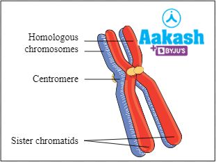
Fig: Structure of chromosome
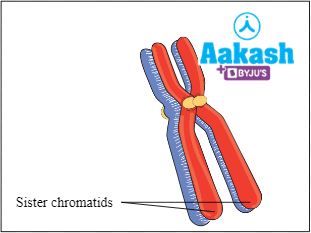
Chromatids
During the prophase stage of mitosis, each chromosome duplicates and forms two sister chromatids that are connected by a centromere during cell division. The sister chromatids are identical to each other and they divide into individual chromosomes during anaphase.
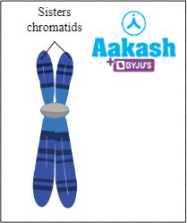
Fig: Chromatids
Centromere
The central portion of a chromosome is called centromere. The centromere links the two chromatids together. Centromere divides the chromosome into short p arm and long q arm. It is also called primary constriction.
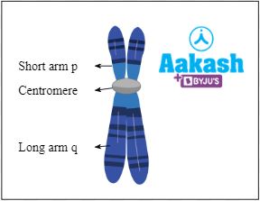
Fig: Centromere
Kinetochore
Proteinaceous disc shaped structure seen on either side of the centromere is called kinetochore. It helps in cell division. Its role is to allow chromosomal mobility during the anaphase stage of cell division.

Fig: Kinetochore
Telomere
The terminal end of the chromatids are called telomeres.
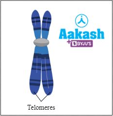
Fig: Telomeres
Secondary constriction
Apart from the primary constriction, some metaphase chromosomes may have one or more than one secondary constriction. It can be found in sites other than primary constriction, mostly at terminal ends. Secondary constrictions are considered as narrow areas of two types such as joints and NOR (Nucleolar Organiser Region). Joints are mainly involved in breaking and fusion of chromosome segments. NOR, as the name suggests, is responsible for the formation of nucleolus. Secondary constrictions can serve as markers because they are consistently present in the same locations.
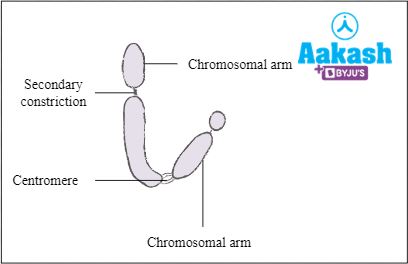
Fig: Chromosomes with secondary constriction
Satellite
Part of the chromosome separated by secondary constriction is called satellite. Chromosomes having satellites are also known as SAT chromosomes. Satellites help in distinguishing between organisms.
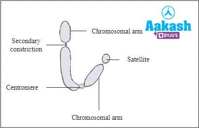
Fig: Chromosomes with a satellite
Matrix
The membrane enclosing each chromosome is known as a pellicle. The jelly-like substance found inside the pellicle is called matrix. It is made of non-genetic materials such as RNA, proteins and lipids.
Classification of chromosomes
When the site of the primary constriction or centromere is clearly apparent during metaphase and anaphase, the chromosomal shape is typically visible. The two arms are equal in iso brachial chromosomes and unequal in the heterobrachial chromosome. The ratio between two arms of a chromosome is known as the centromeric ratio. Chromosomes can be classified into four types based on the position of the centromere as follows:
- Metacentric
- Submetacentric
- Acrocentric
- Telocentric
Metacentric chromosomes
These chromosomes have the centromere at the centre with two equal arms. Here the p arm = q arm.
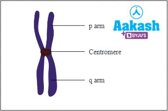
Fig: Metacentric chromosome
Sub metacentric chromosomes
These chromosomes have the centromere near the centre or they have a subterminal centromere with one shorter arm and one longer arm. Here the p arm < q arm.
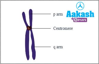
Fig: Submetacentric Chromosome
Acrocentric chromosomes
These chromosomes have the centromere close to its ends forming one extremely short and one very long arm. Here the p arm < < q arm.
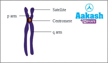
Fig: Acrocentric chromosome
Telocentric chromosomes
These chromosomes have terminal centromeres. Here there is no p arm, only q arm is present.
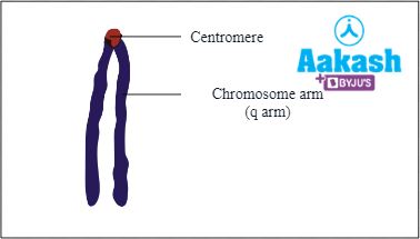
Fig: Telocentric chromosome
Chromatids
A chromatid is considered one-half of a replicated chromosome. Chromosomes get copied and make identical twins before cell divisions. These copies are joined at their centromeres. The joined strands here are called sister chromatids and they are genetically identical. Each strand is called a chromatid. Sister chromatids separate from each other during the anaphase stage of mitosis. At this stage, each separated chromosome is known as the daughter chromosome.
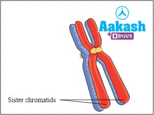
Fig: Chromatids
Fate of sister chromatids in mitosis
During mitosis, each chromosome splits up from the centromere and moves towards the opposite poles. In this way each daughter cell gets a sister chromatid. This can also be called a daughter chromosome. This process helps in the formation of two distinct but identical cells.
GIF: Fate of sister chromatids in mitosis
Fate of homologous chromosomes in meiosis
During meiosis, homologous pairs of parental chromosomes (maternal and paternal copies) meet at the metaphase plate. Each pair here is called bivalent or tetrad. They move away from each other and reach the opposite poles. This will result in the formation of two daughter cells.
GIF: Fate of homologous chromosomes in meiosis
Exchange of genetic material occurs when sister chromatids come closer to each other. It is called the sister-chromatid exchange or SCE. It is the process where two sister chromatids break and rejoin with one another by physically exchanging regions of the parental strands in the duplicated chromosomes.
GIF: Exchange of genetic material
Nondisjunction
Sometimes sister chromatids in mitosis or homologous chromosomes (maternal and paternal copies) in meiosis will not get separated properly. This condition is called a nondisjunction. This condition can occur in the anaphase and can lead to unequal distribution of chromosomes in the daughter cells. It will lead to genetic diseases. Examples include Down’s syndrome, Klinefelter's syndrome etc.
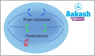
Fig: Nondisjunction
Difference between chromosome and chromatids
The following are the major differences between chromosomes and chromatids:
Chromosome |
Chromatid |
|
The primary function of the chromosome is to transfer genetic material from parents to offspring |
The primary function of the chromatid is to help the cell by duplicating during cell division |
|
It consists of a single, double stranded DNA molecule |
It consists of two DNA strands joined together by centromere |
|
It is the most condensed form of DNA |
It is part of chromosome |
|
Homologous chromosomes are not identical |
Homologous sister chromatids are identical |
|
It appears in M phase |
It appears in interphase |
|
It is thin and ribbon like structure |
It is thin and long structure |
|
A chromosome occurs throughout the cell’s life cycle |
A chromatid is created only when the cell passes through mitosis or meiosis stages of cell division |
|
|
|

Practice Problems
1. The structural element of chromatin is _____________.
- histone
- nucleosome
(C) acid protein and DNA
(D) nuclear matrix
Solution: Chromatin is a protein-DNA complex found inside the nucleus of eukaryotic cells. The nucleosome is the structural unit of chromatin. The negatively charged DNA strand in the cells wraps tightly around the histone molecules (octamer core) to form a nucleosome. It typically has around 200 base pairs of DNA helix. The nucleosomes form a beads-on-a-string structure which represents chromatin. Hence the correct option is (B).
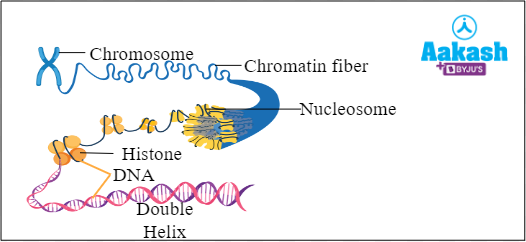
Fig: Condensation of chromatin to form chromosome
2. Chromosome carrying centromeres at one end is ___________________.
- Metacentric
- Submetacentric
(C) Acrocentric
(D) Telocentric
Solution: Chromosomes are rod-shaped structures made up of condensed chromatin fibres. The chromosomes are prominent during the metaphase of the cell cycle. They are responsible for the storage and transmission of genetic information to the daughter cells. Centromere is the region of the chromosome where the two chromatids are joined. On the basis of the position of the centromere, chromosomes are of four different types such as metacentric chromosomes, submetacentric chromosomes, acrocentric chromosomes and telocentric chromosomes.
In telocentric chromosomes, the centromere is found in the terminal position and is capped by the telomere. Hence, the correct option is (D).
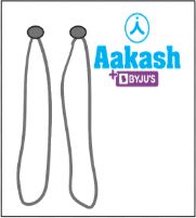
Fig: Telocentric chromosome
3. What is the SAT Chromosome?
Answer: Part of the chromosome separated by secondary constriction is called a satellite. Chromosomes having satellites are also known as SAT chromosomes.
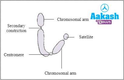
Fig: Chromosomes with a satellite
4. What is meant by kinetochore?
Answer: Proteinaceous disc shaped structure seen on either side of the centromere is called kinetochore. It helps in cell division. Its role is to allow chromosomal mobility during the anaphase stage of cell division.
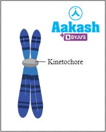
Fig: Kinetochore
FAQs
1. What is meant by karyotype?
Answer: Karyotype represents the complete set of chromosomes (at metaphase stage), in any cell of a species or an individual organism, that are arranged according to length, centromere location and other traits.
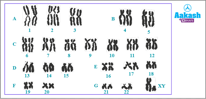
Fig: Human karyotype
2. What are polytene chromosomes?
Answer: C.G. Balbiani discovered polytene chromosomes in the salivary glands of Drosophila melanogaster or fruit fly in 1881. Polytene chromosomes are found in the cells of larvae of Drosophila, mosquitoes and midges and are large chromosomes consisting of thousands of DNA strands. Large chromosomes in polytenes are a result of their high DNA content. They are created through multiple rounds of chromosomal DNA replication during interphase without nuclear division. The resulting daughter chromatids do not split apart and remain joined together instead. The larval cells die during metamorphosis because they are unable to go through mitosis.
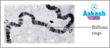
Fig: Polytene chromosome
3. Which are the 2 main types of chromosomes?
Answer: There are two basic types of chromosomes present such as sex chromosomes (allosomes) and autosomes (somatic chromosomes). Autosomes control the inheritance of all the characteristics except the sex-linked charcteristics. These are controlled by the allosomes or sex chromosomes. For example, humans possess 22 pairs of autosomes and one pair of sex chromosomes (XX in females and XY in males).
4. Which is considered as the biggest chromosome in human beings?
Answer: Chromosome I is the largest chromosome in human beings. The chromosome I possess 2968 genes.