-
Call Now
1800-102-2727

Small Intestine: Structure and Role in Digestion, Absorption in Small Intestine, Practice Problems and FAQs

All living organisms require energy to perform work. You know that this energy comes from the food they eat. For example, you can see how this frog, let's call him ‘Babloo’ catch butterfly, it is his food.
GIF: Capturing food
But do you know how the food you eat provides energy? What happens to the ingested food?
What all structures and processes are involved in the digestion of food?
The food we eat first gets digested with the help of various structures such as buccal cavity, oesophagus, stomach, small intestine and large intestine present in the alimentary canal. After digestion, the food is absorbed into the blood and lymph capillaries from where it is assimilated to the various tissues and organs and provides energy.
The small intestine is the major site where most of the absorption of nutrients takes place. Let’s understand more about the small intestine in this article.
Table of contents:
- Structure of small intestine
- Digestion in small intestine
- Absorption in small intestine
- Practice Problems
- FAQs
Structure of Small Intestine
The small intestine is a tube-like structure which is long, narrow and coiled. The structure of the small intestine is divided into three parts, duodenum, jejunum, and ileum.
Duodenum
Duodenum is considered as the shortest and widest part of the small intestine. It is a C-shaped or U-shaped structure.
Jejunum
Jejunum is considered as the middle part of the small intestine. It is a coiled tube with a diameter of about 4 cm and 2.5 cm long. It is a muscular part and therefore, it is reddish in appearance.
Ileum
Ileum is the longest part of the small intestine. It is a highly coiled structure with a diameter of 3.5 cm and 3.5 m long. The wall of the ileum is thinner as compared to the wall of the jejunum.
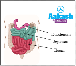
Fig: Small intestine
Peyer’s patches
Peyer patches are a unique feature of the ileum that are named after Johann Conrad Peyer. These are specialised clusters of the lymphatic tissue. The lymphatic tissue produces lymphocytes.
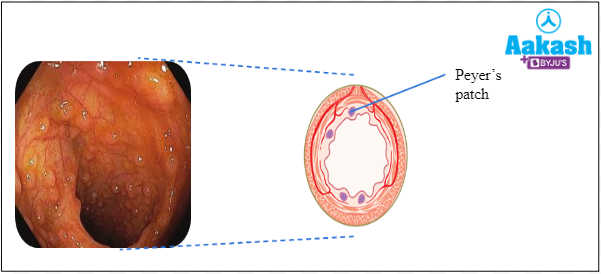
Fig: Peyer’s patches
Plicae circulares
These are one of the prominent structures in jejunum. Plicae circulares are circular folds of the mucous membrane and also known as valves of Kerckring. They increase the surface area for absorption.
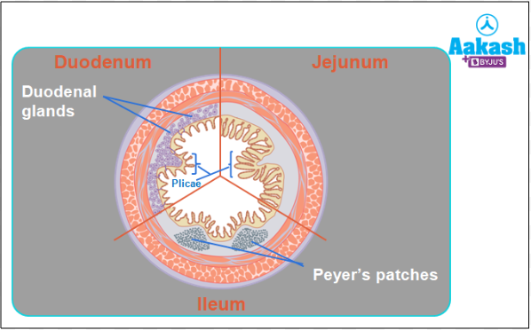
Fig: Structures in the small intestine
Villi
Intestinal villi are the finger-like projections that increase the surface area for absorption.
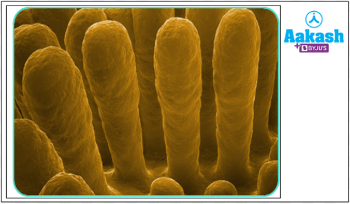
Fig: Villi in small intestine
Intestinal villi is made up of mucosa layers. It increases surface area for absorption. It is covered by epithelium. Villi have lacteal (a lymph vessel) and blood capillaries (vein and artery).
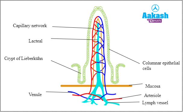
Fig: Structure of villi
Microvilli
Microvilli are microscopic projections present in a single villus. They collectively give a brush border appearance. They increase the surface area for absorption of food. These structures are absent on Peyer’s patches. Blood vessels and lacteals are situated close to the microvilli. The nutrients from the microvilli are transported to the blood vessels and lacteals.
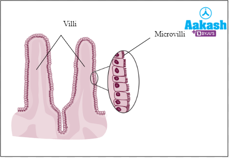
Fig: Structure of microvilli
Digestion in small intestine
Chyme enters the small intestine through the pyloric sphincter.
Mechanical digestion
The automatic movement of the walls of the small intestine (peristalsis) allows the chyme to mix thoroughly with the secretions of the intestine.
Chemical digestion
It occurs with the help of enzymes present or secreted in the small intestine. It occurs with the help of hepato-pancreatic juices and intestinal juices.
Secretions enters duodenum
Secretions that enter duodenum include the following:
- Pancreatic juice
- Bile
- Intestinal juice
Enzymes involved in digestion
There are four main enzymes that are involved in digestion are as follows:
- Carbohydrases secreted by salivary glands, pancreas and small intestine.
- Proteases secreted by stomach, pancreas and small intestine.
- Lipases secreted by pancreas and small intestine.
- Nucleases secreted by pancreas and small intestine.
Carbohydrases
Carbohydrases catalyse the breakdown or hydrolysis of polysaccharides into monosaccharides. This enzyme is produced by the salivary glands, pancreas and the small intestine. The breakdown of polysaccharides takes place in two steps:
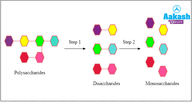
Fig: Hydrolysis of polysaccharides
Proteases
Proteases catalyse the hydrolysis or breakdown of proteins or polypeptides into amino acids. This enzyme is produced by the stomach, pancreas and the small intestine. Protein breakdown takes place in three steps:
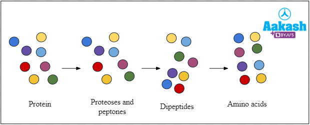
Fig: Hydrolysis of protein
Lipases
Lipases are enzymes that catalyse the hydrolysis or breakdown of fats into fatty acids and glycerol. These are produced by the pancreas and the small intestine. The breakdown of fats happens in three steps as follows:
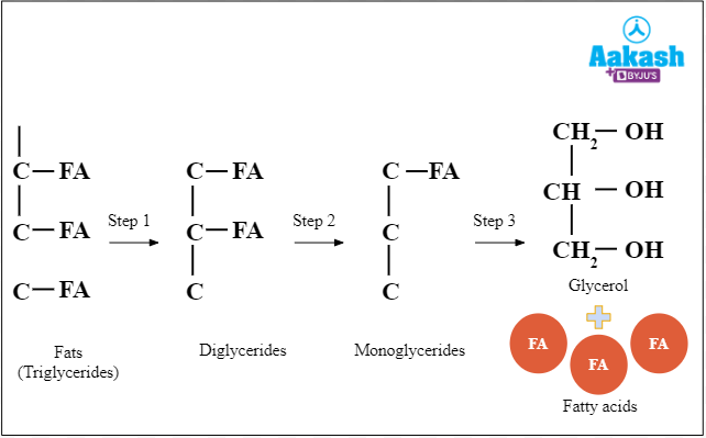
Fig: Hydrolysis of fats
Nucleases
Nucleases are the set of enzymes that catalyse the hydrolysis or breakdown of nucleic acids like RNA and DNA into its components like sugars and bases. This enzyme is produced by the pancreas and the small intestine. The breakdown of nucleic acid takes place in three steps as follows:
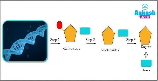
Fig: Hydrolysis of nucleic acid
Pancreatic juice
Pancreatic juice is secreted by the exocrine part called pancreatic acini of the pancreas. It possesses water, bicarbonates and enzymes. It possess the following inactive enzymes:
- Inactive amylases
- Trypsinogen
- Chymotrypsinogen
- Procarboxypeptidase
- Lipases
- Nucleases
Activation of pancreatic enzymes
Activation of trypsinogen
The inactive trypsinogen is converted into trypsin by the action of enzyme enterokinase. It is secreted by cells of duodenum and components of intestinal juice.
Activation of other major pancreatic enzymes
Trypsin activates inactive pancreatic enzymes. Inactive amylases convert into active amylases, chymotrypsinogen converts into chymotrypsin, procarboxypeptidase is converted into carboxypeptidases.
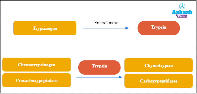
Fig: Activation of major pancreatic enzymes
Bile
Bile is another component which reaches the duodenum. It does not contain any enzyme. It aids in the emulsification of fats.
Components of bile
The components of bile include bile pigments, water, bile salts, and fats. Bile pigments include bilirubin and biliverdin. Bilirubin is orange yellow in colour, whereas the biliverdin is green in colour and it is an oxidised form of bilirubin. Bile salts include sodium glycocholate, potassium taurocholate, potassium glycocholate and sodium taurocholate. Fats include cholesterol and phospholipids.
Intestinal juice
Intestinal juice or succus entericus is secreted by the intestinal glands of Crypts of Lieberkuhn. It contains the following enzymes:
- Disaccharides
- Dipeptides
- Lipases
- Enterokinase
- Nucleotidases
- Nucleosidases
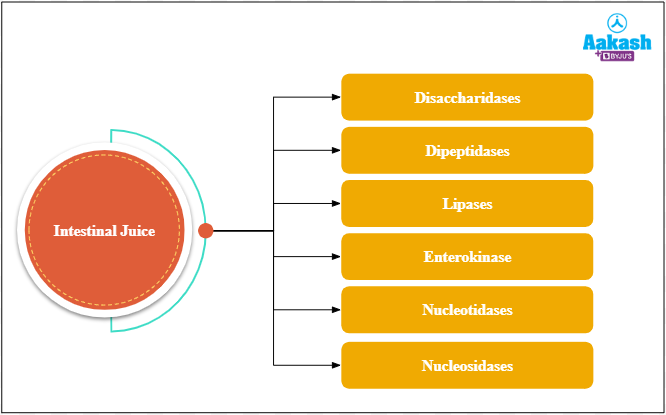
Fig: Enzymes in intestinal juices
Digestion by pancreatic juice
|
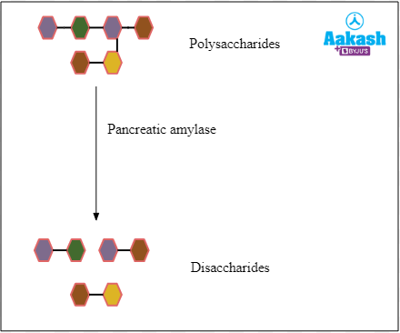
Carbohydrates |
The remaining 70% of the carbohydrates are broken down into disaccharides by pancreatic amylase.
Fig: Hydrolysis of polysaccharides |
|
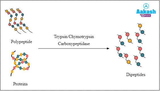
Proteins |
The polypeptides and proteins are digested into dipeptides with the help of trypsin, chymotrypsin and carboxypeptidase.
Fig: Hydrolysis of proteins |
|
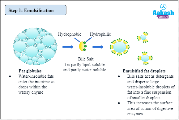
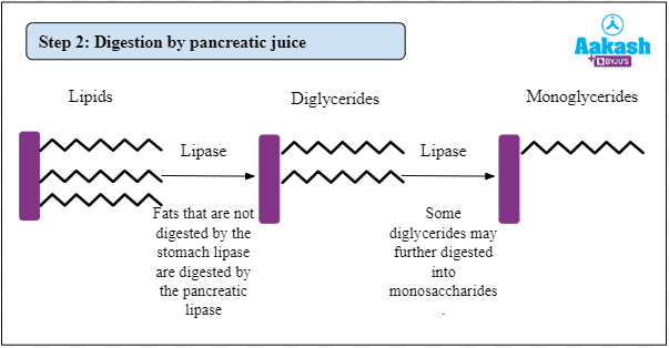
Lipids |
Digestion of lipids by the pancreatic juices occurs after emulsification by bile.
Fig: Emulsification of fat molecules
Fig: Hydrolysis of lipids |
|
Nucleic acids |
Nucleic acids are broken down with the help of nuclease enzymes in the pancreatic juice.
Fig: Hydrolysis of nucleic acids |
Digestion by intestinal juice
|
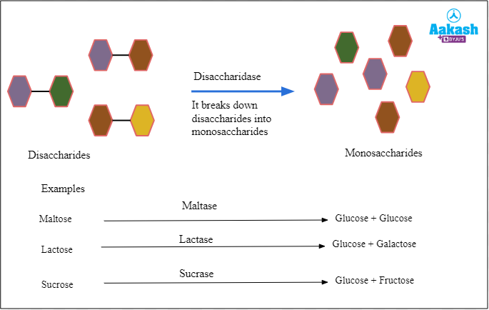
Carbohydrates |
Disaccharides are broken down into monosaccharides by the action of disaccharidases. For example, maltose is broken down by maltase into glucose units.
Fig: Hydrolysis of disaccharides |
|
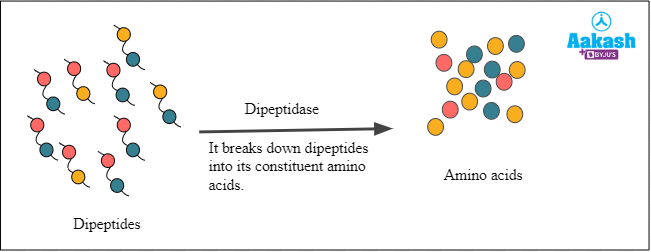
Proteins |
The dipeptides are broken down into constituent amino acids with the help of dipeptidase enzyme.
Fig: Hydrolysis of dipeptides |
|
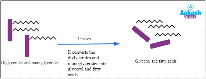
Lipids |
Lipases convert diglycerides and monoglycerides into glycerol and fatty acids.
Fig: Hydrolysis of diglycerides |
|
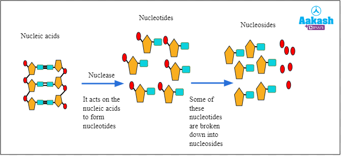
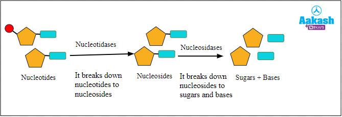
Nucleic acids |
The nucleotides are broken down into nucleosides with the help of nucleotidases. Then nucleosides are broken down into sugars and bases with the help of nucleosidases.
Fig: Hydrolysis of nucleotides |
Absorption in the small intestine
Most of the absorption of nutrients takes place in the small intestine. The nutrients absorbed in the small intestine include glycerol, fatty acids, fructose, glucose and amino acids. The absorption in the small intestine takes place with the help of villi and microvilli.
Major mode of absorption of nutrients in the small intestine
Different types of nutrients are absorbed through different mechanisms in the small intestine. These are discussed below:
|
Nutrients |
Transportation from the intestinal lumen to the intestinal epithelial cells |
Transportation from the intestinal cells to the blood vessels or lacteal |
|
|
Carbohydrates |
Fructose |
Facilitated diffusion |
Facilitated diffusion into the blood vessels |
|
Glucose Galactose |
Active transport coupled with transport of sodium ions |
Facilitated diffusion into the blood vessels |
|
|
Amino acids |
Active transport with the transport of sodium ions |
Simple diffusion |
|
|
Fatty acids and glycerol |
In the lumen, lipid droplets (containing fatty acids and glycerol) combine with bile salts to form micelles. Micelles normally enter epithelial cells by simple diffusion method |
Inside the cells, the micelles form small protein-coated fat globules known as chylomicrons, which are released into the lacteals |
|
Practice Problems
Q1. Identify the incorrect statement regarding the small intestine.
A. Churn and mix ingested food
B. Absorb glycerol, fatty acid, glucose and amino acid
C. Villi and microvilli are present
D. Secrete salivary amylase
Solution: Intestinal villi are the finger-like projections that increase the surface area for absorption. Microvilli are microscopic projections present in a single villus. They collectively give a brush border appearance. Most of the absorption of nutrients takes place in the small intestine. The nutrients absorbed in the small intestine include glycerol, fatty acids, fructose, glucose and amino acid. The absorption in the small intestine takes place due to the presence of villi and microvilli.
Hence, the correct option is d.
Q2. Determine which is not a structural part of the small intestine.
A. Ileum
B. Ischium
C. Jejunum
D. Duodenum
Solution: Small intestine is a tube-like structure which is long, narrow and coiled. The structure of the small intestine is divided into three parts, duodenum, jejunum, and ileum. Ischium is a part of the hip bone which is not related to the small intestine. Hence, the correct option is b.
Q3. The epithelial cells of the small intestine have ___________ on their surface which helps in the absorption of food.
A. Pinocytic vesicles
B. Microvilli
C. Zymogen granules
D. Phagocytic vesicles
Solution: Microvilli are microscopic projections present in a single villus. They collectively give a brush border appearance. They increase the surface area for absorption of food. These structures are absent on Peyer’s patches. Blood vessels and lacteals are situated close to the microvilli. The nutrients from the microvilli are transported to the blood vessels and lacteals.
Q4. Identify the incorrect statement about jejunum.
A. around 2.5 metres long
B. more vascular
C. middle part of the small intestine
D. C-shaped
Solution: Jejunum is characterised as the middle part of the small intestine. It is a coiled tube with a diameter of about 4 cm and 2.5 cm long. It is a muscular part and therefore, it is reddish in appearance. It is more vascular as compared to the other parts of the small intestine. Hence, the correct option is d.
FAQs
Q1. Why is the small intestine also known as small bowel?
Answer: The small intestine is also known as the small bowel because it runs from the stomach to the large intestine. The small intestine is a tube-like structure which is long, narrow and coiled. The structure of the small intestine is divided into three parts, duodenum, jejunum, and ileum.
Q2. What happens if the cells of the small intestine are not working properly?
Answer: Short bowel syndrome is a condition that usually affects people who have lost most of their small intestine through surgery. Without this particular part, the body cannot get enough nutrients and water from the food we eat. This will result in bowel troubles, like diarrhoea. This can be dangerous if left without treatment.
Q3. What symptoms appear if something is wrong in the small intestine?
Answer: If there is something wrong in the small intestine, a number of symptoms appear, such as abdominal pain, spasms, gas, diarrhoea, constipation etc.
Q4. Name three common disorders of the small intestine.
Answer: The common disorders of the small intestine are celiac disease, bowel obstructions, and Crohn’s disease.