-
Call Now
1800-102-2727

Liver: Location, Structure, Duct system, Functions, Common Disorders, Signs of Damage, Diagnosis, Practice Problems, and FAQs

We all take a variety of food items daily. It provides us with the required nutrients for growth and development. Some of you love to eat junk foods loaded with cheese and butter, right?. For example, most of you enjoy eating deep fried food items like pooris, bhature, sweets and fries. Some of you also love ghee on rotis. Ghee not only makes sour rotis soft to eat but also enhances the taste. But have you ever thought, how such heavy food items with oils and fats get digested in our body?
Fig: Fried food items

The cells in our body perform various types of metabolic activities throughout day and night. They produce a lot of harmful wastes as byproducts. How does our body get rid of these wastes? The answer to all these questions lies in the efficiency of the wonderful organ called the liver. So come, let’s discuss more about the liver in this article.
Table of contents
- Liver
- Location of liver
- Structure of liver
- Duct system of liver
- Functions of liver
- Common disorders of liver
- Signs of liver damage
- Diagnosis of a damaged liver
- Practice Problems
- FAQs
Liver
Liver is a bilobed organ. The word liver originated from the Greek word ‘hepar’. It is a vital organ which means one can not survive if the liver malfunctions or removal of the entire liver leads to death. It is the largest gland in the human body. It weighs around 1.3 to 1.5 kgs in a normal healthy adult.
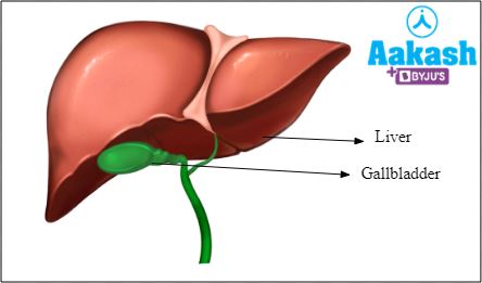
Fig: Liver
Location of liver
It is located in the upper right corner of the abdominal cavity, just below the diaphragm. It is because of the presence of the liver, the right lung is positioned slightly above as compared to the level of the left lung and the right kidney is situated slightly lower as compared to the level of the left kidney.
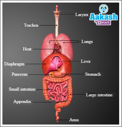
Fig: Location of liver
Structure of liver
The liver has two lobes such as the right lobe and left lobe. Right lobe is larger than the left lobe. The two lobes are held together via a falciform ligament. The entire liver is covered by a thin connective tissue sheath called glisson’s capsule.
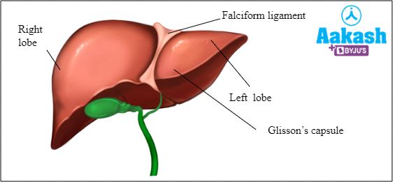
Fig: Structure of liver
Hepatic lobules
The structural and functional units of the liver are called hepatic lobules. These are also covered by glisson’s capsule. The lobules consist of hepatic cells arranged in the form of cords. Each lobule has a polygonal shape (5 to 7 sides).
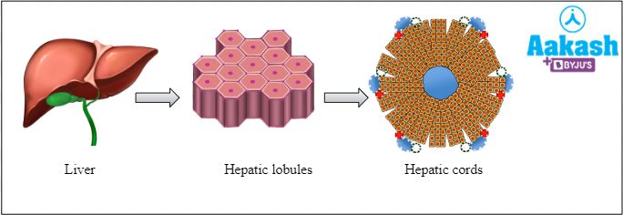
Fig: Hepatic lobules
Anatomy of the hepatic lobule
At the corners of the hepatic lobule polygon, there are portal triads present. Each portal triad has a branch of 2 blood vessels such as a hepatic portal vein, and a hepatic artery, and it also has a branch of the interlobular bile duct. Hepatic artery carries oxygenated blood to the liver cells. Hepatic portal vein brings deoxygenated blood from the organs like the spleen and intestines to the liver. The interlobular bile duct carries bile produced by hepatic lobule. In the centre of the lobule there is a central vein that carries deoxygenated blood away from the lobule and drains into the hepatic vein. The hepatic vein then drains into the inferior vena cava that further carries the deoxygenated blood to the right atrium of the heart. There are kupffer’s cells located in the hepatic lobules which are actually macrophages of the liver.
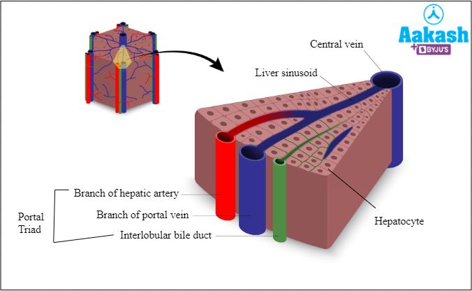
Fig: Anatomy of hepatic lobule
Duct system of liver
The right and left lobes of the liver have their own hepatic ducts, right and left respectively. These two hepatic ducts fuse to form a common hepatic duct. The common hepatic duct carries bile away from the liver to the gallbladder for storage. When required, bile from gallbladder is carried by the cystic duct to outside. This duct receives the common hepatic duct and forms a common bile duct. The bile from liver and gallbladder is carried by the common bile duct towards the duodenum where it also receives a duct from the pancreas called pancreatic duct or Duct of Wirsung. Opening of the common bile duct into the pancreatic duct is guarded by Sphincter of Boyden. Finally, a common hepato-pancreatic duct is formed that opens into the duodenum that drains both, bile and pancreatic juice, into the duodenum. The opening of this duct into the duodenum is guarded by the sphincter of Oddi.
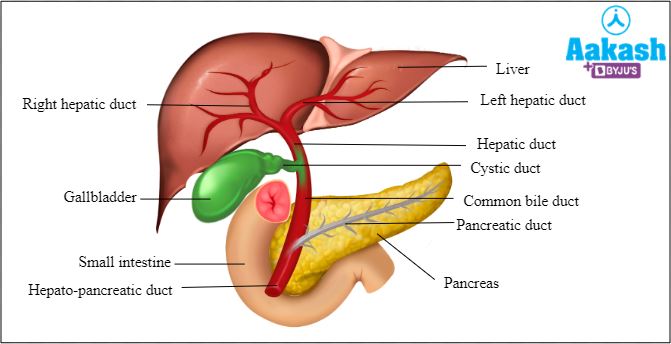
Fig: Duct system of liver,
Functions The following are the major functions of the liver:
- Hepatic portal system
- Detoxification
- Urea formation
- Secretion of bile
- Secretion of heparin
- Excretion
- Pigmentation of faeces
- Metabolism of carbohydrates
- Storage
- Production of plasma proteins
- Processing of haemoglobin
- Erythropoiesis
- Vitamin A synthesis
- Phagocytosis
- Regulation of body heat
Hepatic portal system
The liver forms a special portal circulation with the gastrointestinal tract called the hepatic portal system. This venous system returns blood from the spleen and digestive tract to the liver. Now the raw nutrients in blood are processed before the blood returns to the heart. Excess nutrients are stored in the liver.
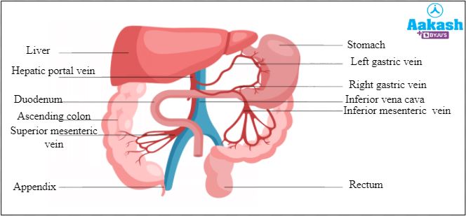
Fig: Hepatic portal system
Detoxification
The harmful metabolites produced by the body cells or man-made chemicals (cigarette smoke, recreational drugs, pollutants, preservatives, etc.) are converted to less harmless substances by the liver.
Urea formation
Ammonia is a highly toxic nitrogenous metabolic waste produced by the cells. It is converted to urea in the liver through the urea cycle (also called Krebs Henseleit cycle or ornithine cycle). Urea cycle is operated in the cytoplasm and mitochondria of hepatic cells. Urea is then removed from the body through the urine produced by the kidney.
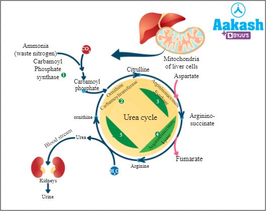
Fig: Urea cycle
Secretion of bile
The liver produces 800 - 1000 ml of bile juice daily. The bile juice aids in digestion of lipids. It is the only digestive juice that does not contain any digestive enzymes. Bile contains bile pigments (bilirubin and biliverdin), bile salts (potassium taurocholate, potassium glycocholate, sodium glycocholate, sodium taurocholate and sodium bicarbonate), phospholipids and cholesterol.
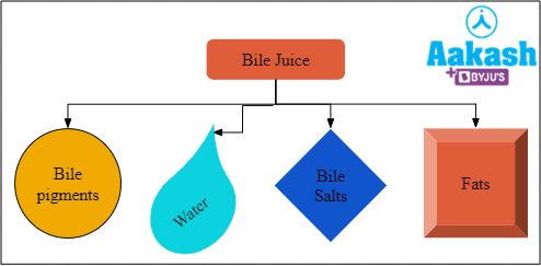
Fig: Components of bile juice
Functions of bile
Bile salts help in emulsification of fats. These salts break down large fat globules into small fat droplets, thereby increasing the surface area for action of pancreatic and intestinal lipase. Lipases help in digestion of lipids and fats.
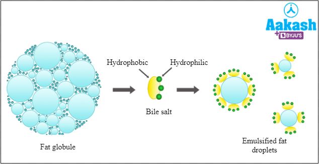
Fig: Emulsification of fats
Secretion of heparin
Heparin is the natural anticoagulant present in blood. It prevents formation of a clot inside the blood vessels and ensures proper blood flow. Heparin is synthesised by mast cells of areolar connective tissue, basophils of blood and liver.
Excretion
Bile juice not only contains bile pigments, bile salts and cholesterol. It also contains degraded steroid hormones, drugs and vitamins. When bile is poured into the duodenum, waste products in bile are eliminated from the body through the anus via the faecal matter. Alcohol is also eliminated from the body by the liver.
Pigmentation of faeces
Bilirubin, the bile pigment, is converted by bacteria to stercobilinogen in the intestine. This stercobilinogen is absorbed and excreted by the kidney or liver. Stercobilinogen is oxidised to stercobilin, which gives colour to faeces.

Fig: Faeces
Metabolism of carbohydrates
Liver is the site of glycogenesis, glycogenolysis and gluconeogenesis. Glycogenesis is formation of glycogen. Glycogenolysis is breakdown of glycogen. Gluconeogenesis is formation of glucose molecules from non-carbohydrate sources like fats and amino acids. Excess glucose is stored in the liver in the form of glycogen by the process of glycogenesis. When required, this glycogen is broken down to release glucose molecules into the blood circulation by the process of glycogenolysis.
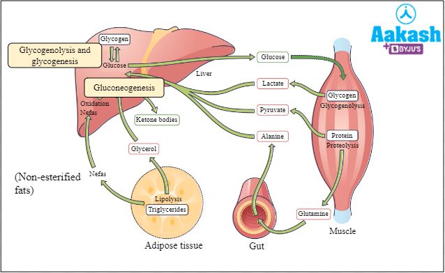
Fig: Metabolism of carbohydrates
Storage
Excess nutrients like glucose and fats are stored in the liver. When required the liver releases them for body tissue utilisation.
Production of plasma proteins
Liver is responsible for secretion of 85 - 90% of circulating protein volume. It includes albumins, globulins and fibrinogens. Most abundant protein in plasma is albumin which is responsible for maintaining osmotic balance.
Processing of haemoglobin
When the RBCs die, the haemoglobin contained in them is broken down in the liver. The globin part being protein, is broken down into amino acids and these are then utilised by cells for production of new proteins. The heme part, the prosthetic group, is separated from iron. Iron is sent to the bone marrow where it is utilised for production of new RBCs and heme is converted to bilirubin.
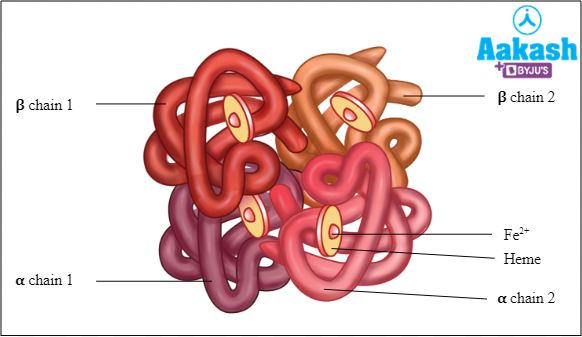
Fig: Structure of haemoglobin
Regulates blood clotting
Liver is the site for synthesis of clotting proteins in their inactive form, like fibrinogen. It is activated to fibrin for clot formation.
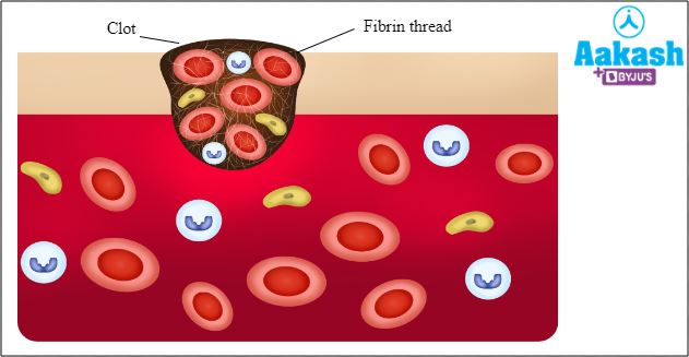
Fig: Formation of blood clot
Erythropoiesis
In early foetal life, RBC formation takes place in the liver.
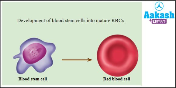
Fig: Erythropoiesis
Vitamin A synthesis
The precursor of vitamin A (retinol) called beta carotene is stored in the liver after being absorbed from the small intestine. As and when required by the body, beta carotene is converted to vitamin A by the stellate liver cells.
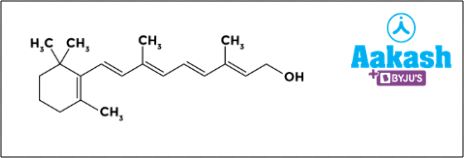
Fig: Retinol
Phagocytosis
The kupffer cells in the liver are phagocytes. They phagocytose any pathogen that infects the liver and protects the liver from infections. They also phagocytose dead cells and foreign matter.
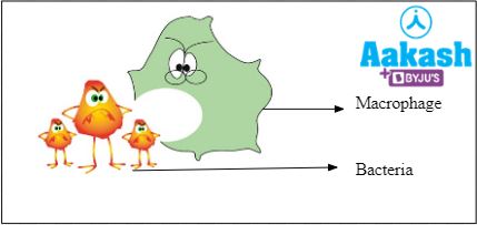
Fig: Phagocytosis
Regulation of body heat
Liver performs a wide range of metabolic activities. Heat is generated as a by-product of these reactions and as the blood passes through the liver it carries this heat and distributes to all parts of the body. This helps in the regulation of body heat.
Common disorders associated with liver
The following are some common disorders associated with liver:
Jaundice
In this condition, the concentration of bilirubin increases in the blood, causing yellowness to skin and eyes. It affects the liver mainly.
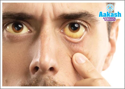
Fig: Jaundice
Fatty liver
This condition is described by excess buildup of fat in the liver. It is also called hepatic steatosis.
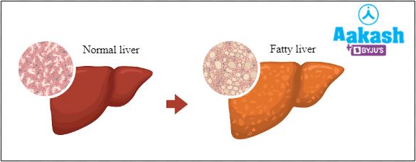
Fig: A healthy vs fatty liver
Hepatitis
Inflammation of the liver by hepatitis viruses or any autoimmune condition is called hepatitis.
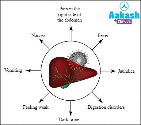
Fig: Common symptoms of hepatitis
Liver failure
In this condition the liver loses its function. This can happen due to medications, high doses of paracetamol, hepatitis, advanced fatty liver or long-term alcohol consumption..
Liver cirrhosis
Long term liver damage can lead to scarring of liver and liver failure. It is called liver or hepatic cirrhosis.
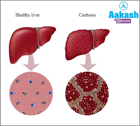
Fig: A normal vs cirrhosis liver
Wilson disease
It is an inherited disease leading to accumulation of too much copper in vital organs like the brain or liver. If left undiagnosed and untreated it leads to liver cirrhosis and death.
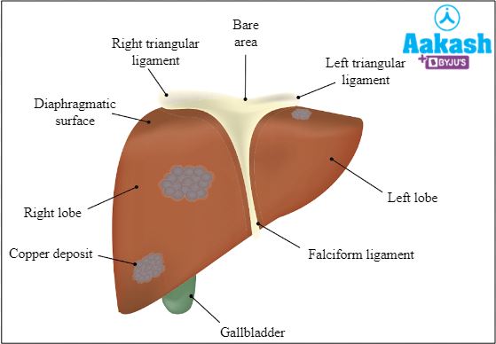
Fig: Wilson disease
Hemochromatosis
It is also an inherited disease which leads to iron overload in the body. It can cause liver cancer, liver cirrhosis, or irregular heart rhythms.
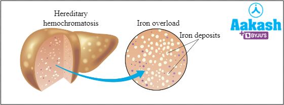
Fig: Hemochromatosis
Common signs of liver damage
Any person suffering from liver damage may show the following signs:
- Abdominal pain and swelling.
- Fluid retention in lower body parts.
- Loss of appetite.
- Nausea and vomiting.
- Dark coloured urine and stools.
- Itchy skin.
- Fatigue
- Weakness
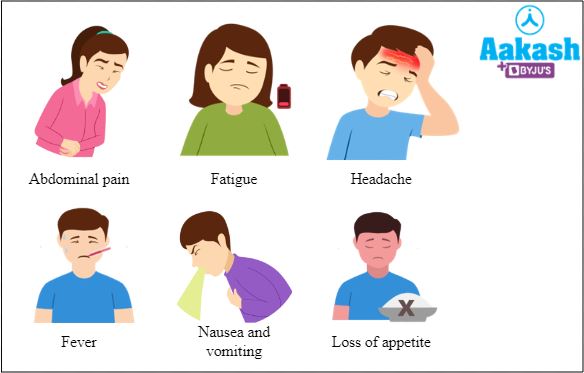
Fig: Common signs of liver damage
Diagnosis of a damaged liver
Any kind of liver damage can be diagnosed by the following tests:
Blood test
A blood test for all liver enzymes and their levels help in analysing any liver damage.
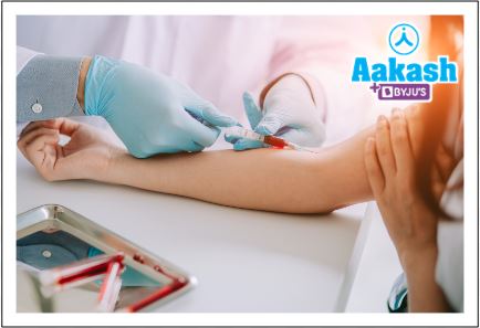
Fig:Blood test
Imaging tests
Liver conditions like fatty liver, tumours, cancer, or cirrhosis can be diagnosed with the help of imaging tests like ultrasound, CT scans or MRI.
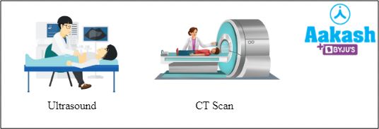
Fig: Ultrasound and CT scan
Liver biopsy
Sometimes a liver biopsy may be required to better understand the conditions like liver tumours and cancer better. In liver biopsy a small piece of liver tissue is removed and examined under a microscope to check the signs of disease or damage.
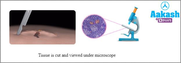
Fig: Biopsy
Practice Problems
Q1: Which of the following organs secretes angiotensinogen?
a. Liver
b. Kidney
c. Lungs
d. Adrenal gland
Solution: Angiotensinogen is a component of the renin-angiotensin-aldosterone system (RAAS) that operates to regulate the body fluid volume. This system is activated when the body fluid volume decreases. Angiotensinogen released by the liver is converted to angiotensin I by renin produced by the juxta glomerular cells (JGA) of the kidney. Angiotensin I is then converted into angiotensin II by angiotensin converting enzymes (ACE) secreted by lungs. Angiotensin II is then responsible for bringing the body fluid volume back to normal. Hence, the correct option is a.
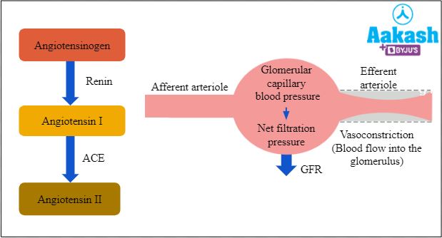
Fig: RAAS mechanism
Q2: If the liver is removed, which of the following nutrients will not be absorbed in the mucosa of the small intestine?
a. Proteins
b. Vitamins
c. Collagen
d. Fats
Solution: Liver secretes bile juice which is responsible for emulsification of fats. It thus helps in digestion of fats in our diet in the small intestine. If the liver is removed, bile juice will not be produced and hence the digestion of fats will get disturbed in the intestine. Hence, the correct option is d.
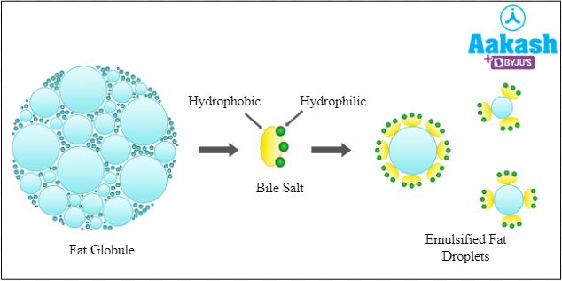
Fig: Emulsification of fats
Q3: Stool of a person is dark brown to black in colour due to malfunction of which organ?
a. Pancreas
b. Kidney
c. Spleen
d. Liver
Solution: Malfunction of liver leads to change in the colour of urine and stool. Hence, the correct option id d.
Fig: Faeces
Q4: Which of the following statements is true for bile juice?
a. It contains enzymes like lipases and amylases
b. It contains only bile pigments and bile salts
c. It contains bilirubin, biliverdin and amylases
d. It does not contain any digestive enzyme
Solution: Bile juice is secreted by the liver. Bile juice is the only digestive juice that does not contain any digestive enzymes. Bile contains bilirubin, biliverdin, bile salts, phospholipids, cholesterol, vitamins, and degraded steroid hormones. Hence, the correct option is d.
FAQs
Q1: How can a person live after donating their liver?
Answer: Liver is an organ that can regenerate. Whenever a person donates their liver, only a part of the liver is donated and not the complete organ. Thus, the remaining part regenerates to the whole liver in the donor. The transplanted liver tissue in the recipient also regenerates to a complete liver.
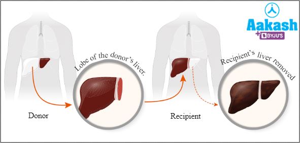
Fig: Liver transplantation
Q2: What happens if gallbladder is removed?
Answer: Surgical removal of gallbladder is called cholecystectomy. It is done if the patient has gallbladder stones that cause significant pain and other complications. After gallbladder removal such patients are advised to cut down significantly on fatty food items like, fatty meat, deep fried items, ghee, butter, cheese, etc.
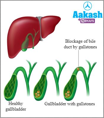
Fig: Healthy vs gallbladder with gallstones
Q3: Are there different types of hepatitis?
Answer: Hepatitis refers to inflammation of the liver. It can be of the following types:
- Hepatitis A - It is caused by hepatitis A virus which spreads through contaminated food and water.
- Hepatitis B - It is caused by hepatitis B virus. This virus is transmitted through sexual contact or from infected needles or body fluids (semen or blood).
- Hepatitis C- It is caused by hepatitis C virus that is transmitted just like hepatitis B virus.
- Hepatitis D - It is caused by hepatitis D virus. It spreads just like hepatitis B and C viruses.
- Hepatitis E - It is caused by hepatitis E virus. It spreads via the faeco-oral route through contaminated food and water.
Q4: How does the gallbladder release the stored bile?
Answer: Liver produces bile juice continuously, irrespective of the presence or absence of lipids and fats in the alimentary canal. To prevent wastage of this bile juice, it is stored in the gallbladder where bile is also concentrated. When we eat fat rich food, a hormone called cholecystokinin is released by the endocrine cells of the small intestine (duodenum). This hormone acts on the gallbladder and causes it to contract which leads to release of bile from the gallbladder into the cystic duct.
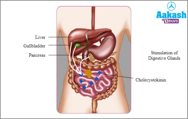
Fig: Secretion of cholecystokinin