-
Call Now
1800-102-2727

Human Skull, Practice Problems and FAQs

The shape of the head varies hugely from one species of animal to another, right? Even within a particular species, say for humans, the shape of the face and the head varies from one person to another, right? Why do you think this happens?
We know that the shape of the body of all vertebrate animals is determined by the endoskeleton which is mainly made up of bones and cartilages. It not only helps to maintain the form and structure of the body but also aids in locomotion. Needless to say that a certain part of the endoskeleton is responsible for the shape of the head and the face too. Can you name this part? Yes, it is the skull. If we compare the skull of a human and the skull of a bull, can we find any differences? Of course, there are differences! Even though humans and bulls are mammals they belong to different species. Consequently, they have different anatomical features such as size of organs, shape of organs, etc and hence their endoskeleton, including the skull, will also reflect differences. However, there are also some basic similarities as both humans and bulls belong to the same class, that is, Mammalia.
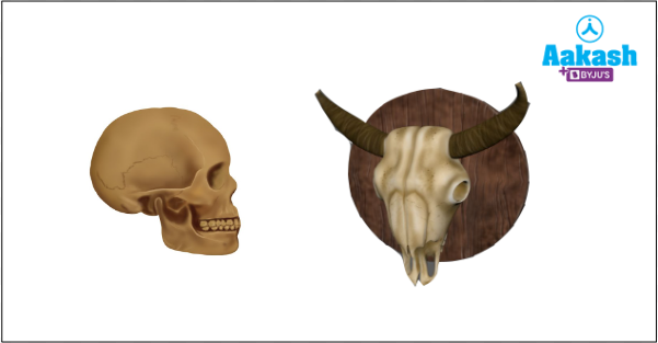
Fig: Skull of human and bull
Now you compare the shape of your face with that of your sibling. You may find some differences in the shape of your head, chin, cheeks, forehead etc. These differences are due to the slight variations in the shape of the bones of your skull. But the types and number of bones present in a human skull will be the same for all humans. But is this all that the skull does? Does it only determine the shape and form of our face and head? Well, no. The main function of the skull is to protect the delicate internal organs in our head from shocks and injuries. How it does so can be understood better only if we study its structure in detail.
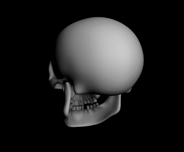
GIF: Human skull
What happens if there are anomalies in the anatomy of a skull? There will obviously be some wide differences in appearance as well as functionality of the skull, and this may occur due to various disorders in the body or some accident and severe injury. In this article we will discuss the structure and function of the human skull and some disorders associated with it. Keep reading!
Table of contents:
- Human skull
- Disorders of human skull
- Significance of human skull
- Practice Problems
- FAQs
Divisions of the human endoskeleton
The human endoskeleton is divided into an axial and an appendicular skeleton. The axial skeleton occupies the longitudinal central axis of the body and is composed of the skull, the vertebral column, sternum and ribs. Out of the 206 bones in the human skeleton, 80 are present in the axial skeleton.
The appendicular skeleton is located on the sides of the axial skeleton and includes the upper and lower limbs and their girdles. It contains 126 bones.
Human skull
The skull is a part of the axial skeletal system. It is the skeleton of a head and possesses 29 bones. The human skull possesses one of the hardest bones in the human body, which is the jawbone .
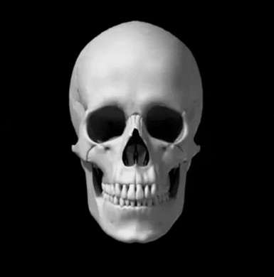
GIF: Human skull
The bones in the skull can be divided into the following regions:
- Neurocranium (8 bones)
- Viscerocranium (14 bones)
- Ear ossicles (6 bones)
- Hyoid (1 bone)
Sometimes hyoid is also considered as the part of viscerocranium.
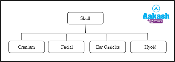
Fig: Parts of a human skull
Neurocranium
The region of the skull which protects the brain is called neurocranium or braincase. It includes the calvaria or skullcap and the remaining bones of the skull comes under the facial skeleton or viscerocranium. Eight bones of the skull comprise neurocranium. There are some zig-zag lines on the cranium, which represent the immovable joints and are known as sutures. Cranial bones are joined together through sutures. The different types of cranial bones are as follows:
- Ethmoid bone (1)
- Frontal bone (1)
- Parietal bone (2)
- Occipital bone (1)
- Temporal bone (2)
- Sphenoid bone (1)
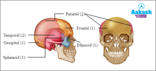
Fig: Neurocranial bones
Ethmoid bone
The unpaired bone in the skull located at the roof of the nasal cavity between the orbits of the eyes is called ethmoid bone. It separates the nasal cavity and the brain. The ethmoid bone can be observed as an irregular bone that makes up the medial wall of orbit of the eye. The three parts of ethmoid bone are as follows:
- Cribriform plate - The roof of the nasal cavity is formed by a cribriform plate.
- Ethmoidal labyrinth - The lateral mass of the ethmoid bone is the ethmoid labyrinth and it is composed of thin walled cellular cavities.
- Perpendicular plate - It is the polygonal, flattened lamina which descends from the lower surface of the cribriform plate. The superior part of the nasal septum is formed by the perpendicular plate of the ethmoid bone.
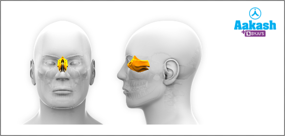
Fig: Ethmoid bone
Frontal bone
The single flat bone that makes the forehead and upper portion of the eye sockets is called frontal bone. The two parts of frontal bone are as follows:
- Squamous part (vertical and biggest part)
- Orbital part (horizontal part)
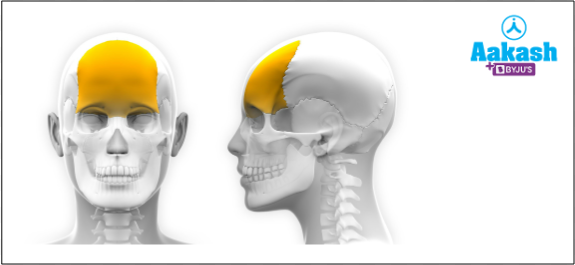
Fig: Frontal bone
Parietal bones
A pair of flat bones that are present on either side of the head or behind the frontal bone is called parietal bones. They join at a fibrous joint and also form the sides and roof of cranium. The outer surface of parietal bones are smooth, but the inner surface has some depression and numerous furrows.
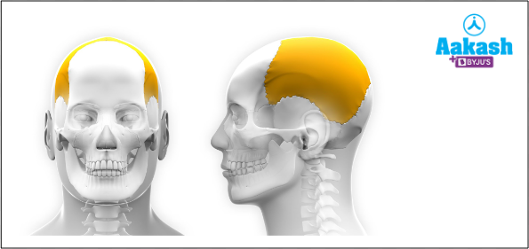
Fig: Parietal bone
Occipital bone
The flat and trapezoidal shaped bone located at the back of the skull is known as the occipital bone. It has an opening called foramen magnum at the base of the skull. There are three main parts for occipital bone as follows:
- Basilar part or basioccipital - This is the part of occipital bone that extends forward and upward from the foramen magnum.
- Lateral parts or ex occipitals - This part is located at the sides of the foramen magnum.
- Squamous part - This is the part of occipital bone that is located above and behind the foramen magnum. This part is curved from above and on the sides too.
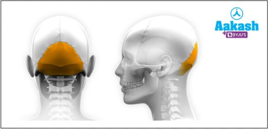
Fig: Occipital bone
Foramen magnum
The large oval opening of the skull where the spinal cord and its accompanying structures pass through is known as foramen magnum. It also passes the accessory nerve into the skull. Foramen magnum is an important part in the bipedal mammals, because the foramen magnum helps these animals to maintain the posture and helps in locomotion. Even though the foramen magnum helps in the bipedal locomotion, the functional correlation is not yet clear.
Occipital condyles
There are two types of convex projections on either side of the foramen magnum that can movably articulate with the atlas vertebra. These are known as the occipital condyles. The human skull is considered as the dicondylic skull as we have two occipital condyles. Even amphibians are known to have dicondylic skulls.
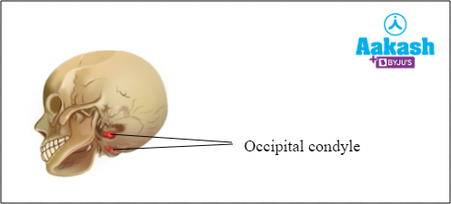
Fig: Dicondylic skull
Animals such as birds and reptiles have a single occipital condyle and are hence said to have monocondylic skulls.
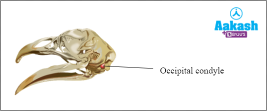
Fig: Monocondylic skull
Temporal bones
A pair of irregular bones that are present lateral to each of the parietal bones and form the sides and base of the skull are called the temporal bones. The major blood vessels of the brain and the lowest seven cranial nerves pass through temporal bones. They protect the temporal bone of the brain and are present around the ear canal. The major four parts of temporal bones are as follows:
- Squamous - It is the largest and superiorly positioned part of the temporal bones which forms the front and upper part.
- Mastoid - It is located in the posteroinferior part of squamous bones and forms the posterior part of the temporal bone.
- Petrous - It is the diamond shaped part of temporal bone fused with the squamous part and mastoid part. It is located between the sphenoid and occipital bones.
- Tympanic - It is the smallest part that lies inferior to the squamous part and anterior to the mastoid part.
There is a long, arched process called the zygomatic process, which projects from the lower region of the squamous part of the temporal bone. It joins with the temporal process of the zygomatic or cheek bone to form the zygomatic arch.

Fig: Temporal bone
Sphenoid bone
The unpaired irregular bone that is situated posterior to the frontal bone and anterior to the occipital bone is called the sphenoid bone. This bone forms the posterior surface of the orbit of the eyes. It forms a connection between the neurocranium and the viscerocranium (facial skeleton) as it articulates with the the frontal, parietal, ethmoid, zygomatic, temporal, occipital, palatine, and vomer bones. The shape of the bone is similar to that of the wings of a butterfly when the bone is extended. The parts of sphenoid bone is as follows:
- Body - contains sella turcica which has a pituitary gland. It also contains paranasal sinuses and the sphenoidal sinuses.
- Two greater wings and two lesser wings - Greater wings are on the lateral sides of the body of sphenoid bone. The lesser wings are on the anterior side.
- Pterygoid process - The part of sphenoid bone that is directed downwards. It is positioned in the junction between the body and greater wings.
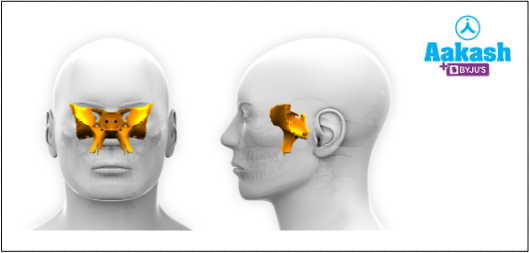
Fig: Sphenoid bone
Viscerocranium
The region of the skull which forms the face is called viscerocranium or facial skeleton. The number of facial bones in the human skull is 14. These bones collectively protect the soft facial tissues. It can also help in other activities like breathing, eating, facial expressions and speech. The different bones included in the viscerocranium are as follows:
- Nasal bones (2)
- Palatine bones (2)
- Lacrimal bones (2)
- Zygomatic bones (2)
- Maxillary bones (2)
- Inferior nasal conchae (2)
- Vomer bone (1)
- Mandible (1)
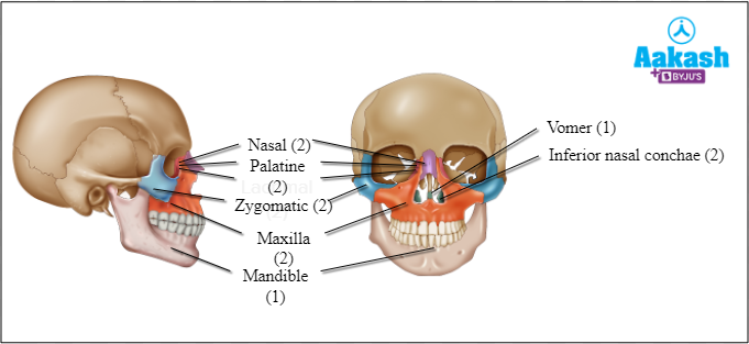
Fig: Facial bones
Nasal bones
The two oblong bones placed side by side at the nasal area are called nasal bones. These bones are of different form and size in different individuals. The two bones form the bridge of the nose and they are joined through a midline internasal suture.
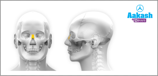
Fig: Nasal bone
Palatine bones
The two irregular ‘L’ shaped bones located above the uvula in the throat are called palatine bones. They comprise the hard palate along with the maxillae. The walls of the three cavities like the floor and lateral walls of the nasal cavity, roof of mouth and the floor of the orbits are formed by the palatine bones.
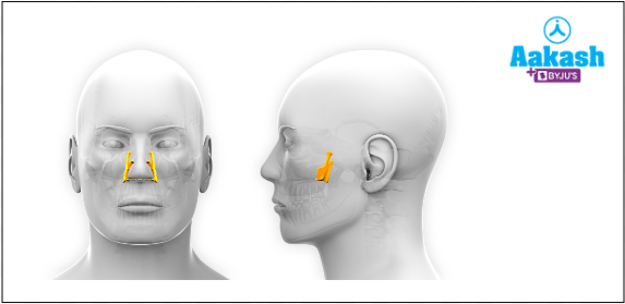
Fig: Palatine bone
Lacrimal bones
The two small fragile bones among the facial bones which are situated at the front part of the medial wall of eye orbit are called the lacrimal bones. Each lacrimal bone is only the size of the little fingernail. It helps in the process of crying or lacrimation by forming the nasolacrimal canal which allows the translocation of tears from the eyes to the nasal cavity. A depression in the anterior part of the lacrimal bone known as the lacrimal fossa consists of the lacrimal sac which stores the excess tears released from the lacrimal glands and allows it to drain into the nasopharynx through the nasolacrimal gland.
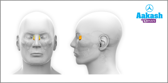
Fig: Lacrimal bone
Zygomatic bones
The facial bones on the upper and lateral part of the face on the cheek area are called zygomatic bones. These are two in number and are also called cheekbone or malar bone. It is also a part of the floor of orbit.
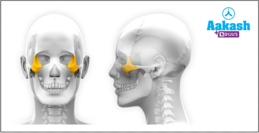
Fig: Zygomatic bone
Maxilla
The fixed bone on the upper part of the jaw is called the maxilla. It is formed by the fusion of two maxillary bones. The upper jaw is composed of the hard palate in the front part of the mouth. Each maxilla attaches to the cheek or zygomatic bones.
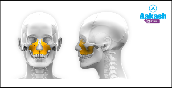
Fig: Maxillae
Inferior nasal conchae
One of the three paired nasal conchae in the nose which extends through the lateral walls of the nasal cavity are called the inferior nasal conchae. It is composed of a lamina of spongy bone and it is shaped like a scroll since it is curled upon itself. The upper and middle nasal conchae arise from the ethmoid bone and are considered to be a part of the neurocranium. The nasal conchae are lined with mucus and help to clean the inhaled air and make it moist and warm as it passes through the nasal cavity.
Vomer bone
One of the unpaired bones of the skull which forms the inferior part of the nasal septum is called vomer bone.
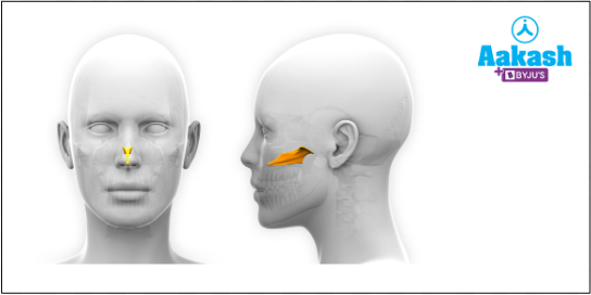
Fig: Vomer bone
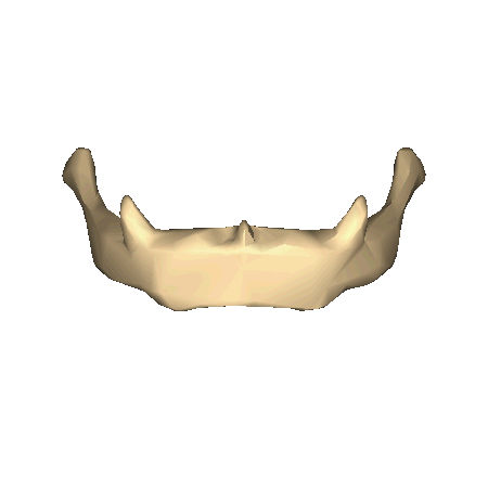
Mandible
The largest and strongest bone of the human facial bones is called the mandible. It is the lower jaw or jaw bone which holds the lower teeth. It is the only bone which is movable in the skull. Usually the mandibles of male are stronger and larger than females.
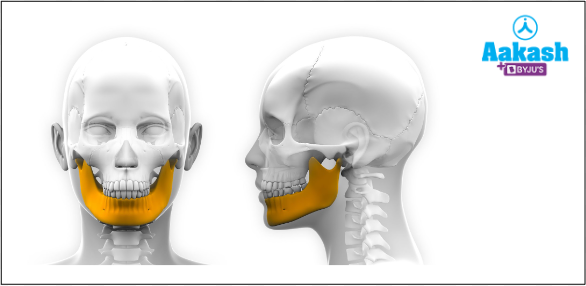
Fig: Mandible
Ear ossicles
The bones present in the middle part of each of the human ear are called ear ossicles. They are also called auditory ossicles. There are three bones present in each ear, hence there are six ear ossicles in a human ear. They are as follows:
- 2 hammer shaped malleus
- 2 anvil shaped incus
- 2 stirrup shaped stapes
The stapes are the smallest bones in the human body.
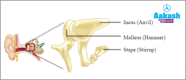
Fig: Ear ossicles
Hyoid bone
The U shaped bone present in the upper part of the throat, above the larynx is called hyoid bone. Certain tongue muscles and the floor of the mouth is attached through hyoid bone.
GIF: Hyoid bone
Disorders of human skull
Most of the disorders that affect the bones also affect the skull. Any abnormalities in the skull will change the shape of the skull along with the shape of the face. If the result of abnormality is the brittleness of the skull bones, then it will affect the protection of internal organs inside the skull. Following are some of the disorders that affect the human skull.
Acromegaly
The excess secretion of growth hormone will result in the abnormal growth of bone tissues in the human body and this condition is known as acromegaly. This condition can cause progressive deformity of the skull through frontal bossing and cranial thickening. This will only occur after the full growth of bones in adults.
Fig: Acromegaly
Paget disease
The condition by which new bone is generated faster than the normal is called paget disease. So this condition interferes with the normal bone recycling process. This will lead to the formation of less organized and fragile bones, which causes pain, deformities and fractures. This condition is usually seen in the pelvis, skull and legs. This condition in the skull causes hearing loss or headaches. The cause for this disease is unknown. According to some scientists, it can be caused due to the combination of genetic and environmental factors.
Fibrous dysplasia
The condition by which a fibrous tissue will develop in the place of normal bone is called fibrous dysplasia. It will make the bone weaken and cause deformities or fracture. It is a genetic disorder which is caused by gene mutation. But this mutation does not pass from parent to child. Along with other bones, it also affects the skull.
Hyperostosis of the skull
The thickening of the frontal bone of the skull is called hyperostosis. The thickening of the skull may be sessile (diffused thickening) or nodular (focal thickening leading to lumps). It is usually seen in women over the age of 65.
Osteoma
The benign growth of new bone tissues over existing bones, typically on the skull is called osteoma. So here a new bone is formed over another piece of bone. If the bone tumor grows on other bones it is known as homoplastic osteoma. It can also grow on other tissues, then it is known as heteroplastic osteoma. This condition can cause headaches and sinus infections.
Significance of human skull
The skull protects all the internal organs present inside it from external damages. Skull also gives shape to the face. The position of the brain is inside the skull, hence the cranium protects the brain from external injuries. There are some sense capsules associated with the cranium. This will help in the protection of eyes, ears and olfactory organs. The jaw bones help to bite and chew food properly. The nasal passages are supported by the skull. Ear ossicles are responsible for the amplification of the sound and clear hearing. The tongue is supported by hyoid apparatus. The facial muscles are attached to the facial bones, such that they help to provide facial expressions.
Practice Problems
1. Which are the wrong statements among the following?
- Skull can be divided into two regions - neurocranium and viscerocranium.
- The zig-zag lines on the cranium, which are immovable, are known as sutures.
- The shape of parietal bone is similar to that of the wings of a butterfly when the bone is extended.
- The stapes is the smallest bone in the human body.
- A and B
- B and C
- Only C
- C and D
Solution: Skull is the skeleton of a head which possesses 29 bones. The skull can be divided into two regions - neurocranium and viscerocranium. The region of the skull which protects the brain is called neurocranium or braincase. Eight bones of the skull comprise neurocranium. There are some zig-zag lines on the cranium, which form the immovable joints and are known as sutures. Cranial bones are joined together through sutures.
A pair of flat bones that are present on either side of the head or behind the frontal bone are called parietal bones. The unpaired irregular bone that is situated posterior to the frontal bone and anterior to the occipital bone is called the sphenoid bone. The shape of this bone is similar to that of the wings of a butterfly when the bone is extended.
The bones present in the middle part of each of the human ear are called ear ossicles. They are also called auditory ossicles. The ear ossicles in each ear are formed of three bones - malleus, incus and stapes. The stapes is the smallest bone in the human body. Hence the correct option is c.
2. Match the following disorders of the brain with their name and condition.
|
Name of the disorder |
Condition |
|
i)Thickening of the frontal bone of the skull |
|
ii) A new bone is formed over another piece of bone |
|
iii) Formation of fibrous tissue in the place of normal bone |
|
iv) Progressive deformity of the skull |
|
v) Formation of less organized and fragile bones |
- A - i, B - ii, C - iii, D - iv, E - v
- A - iv, B - v, C - iii, D - i, E - ii
- A - i, B - ii, C - iv, D - iii, E - v
- A - iv, B - ii, C - iii, D - i, E - v
Solution: The excess secretion of growth hormone in adults will result in the abnormal growth of body tissues in the human body and this condition is known as acromegaly. This condition can cause progressive deformity of the skull through frontal bossing and cranial thickening.
The condition by which new bone is generated faster than the normal is called paget disease. This will lead to the formation of less organized and fragile bones, which causes pain, deformities and fractures.
The condition by which a fibrous tissue will develop in the place of normal bone is called fibrous dysplasia.
The thickening of the frontal bone of the skull is called hyperostosis.
The benign growth of new bones on existing ones, typically on the skull is called osteoma. So here a new bone is formed over another piece of bone. Hence the correct option is b.
3. What is foramen magnum?
Answer: The large oval opening of the skull where the spinal cord and its accompanying structures pass through is known as foramen magnum. It also passes the accessory nerve into the skull. Foramen magnum is an important part in the bipedal mammals, because the foramen magnum helps those animals to maintain the posture and helps in locomotion. However, the functional correlation between the presence of foramen magnum and its role in posture and locomotion of bipeds is not yet clear.
4. What is the type of skull seen in humans?
Answer: Skull can be divided into two types on the basis of the number of occipital condyles on the occipital bones. They are monocondylic and dicondylic. In humans, a dicondylic skull is observed. The skull having two rounded occipital condyles present at the posterior end of the cranium is called the dicondylic skull. These condyles help in articulation with the first vertebrae, the atlas. Dicondylic skulls are usually seen in amphibians and mammals.
FAQs
1. What is magnetoreception?
Answer: The sense of an organism to detect the magnetic field of earth is called magnetoreception. The ethmoid bones, which are a part of cranial bones of organisms like birds and other migratory animals help in magnetoreception. So, the ethmoid bones have the deposits of biological magnetite, which helps to sense the magnetic field. Humans have a vestigial magnetic deposit.
2. What is ossification?
Answer: The process of the formation of a new bone is called ossification. This process begins in the third month of a human fetus. Ossification will be completed by late adolescence. In this process, two types of bones are formed. One is the compact bone, which makes up 80 percent of the skeleton. The second one is the cancellous or spongy bone, which includes the skull, shoulder blades and the ends of long bones.
3. What is a coronal suture?
Answer: A dense and fibrous connective tissue that separates the two parietal bones of the skull from the frontal bones is called coronal suture. Premature fusion of the sutures happens when certain bones of the skull grow faster than normal. This will result in skull deformities.
4. What is Jacobson's organ?
Answer: The chemoreceptor organ which is close to the vomer and nasal bones is called the Jacobson's organ. Hence it has a name, the vomeronasal organ. It is developed in cats which help to perceive certain pheromones.