-
Call Now
1800-102-2727

Microsporogenesis, Structure of Microsporangium or Pollen Sac, Steps Involved in Microsporogenesis, Microgametogenesis, Pollen Structure, Significance of Microsporogenesis, Practice Problems and FAQs

You must be aware that many people suffer from seasonal allergies. Do you know which allergen is responsible for these seasonal allergies? It is the fine, yellowish powdery substance, known as pollen, which is produced by some flowers to carry out sexual reproduction. Pollen is actually a mass of microspores which carry the male gamete in flowering plants. The pollen grains travel from one flower to the female gametes of another flower to carry out the process of fertilisation and sexual reproduction. This process of pollen transfer is known as pollination. Many plants use wind as an agent of pollination during their flowering season and the abundance of pollen in the air often triggers allergies in susceptible people.
Do you know how these pollen are produced? What is the name of the process? What is the site of pollen production? You can find the answers to all these questions in this article. So, let’s dive into it.
Table of contents
- Introduction
- Microsporangium or Pollen sac
- Steps involved in microsporogenesis
- Microspore to pollen
- Microgametogenesis
- Dehiscence of anther to release pollen
- Pollen structure
- Significance of microsporogenesis
- Practice Problems
- FAQs
Introduction
The process through which pollen grains or microspores are formed is known as microsporogenesis. This process takes place inside the pollen sacs or microsporangium of flowering plants. The formation of pollen grains occurs through meiotic or reductional division. Each microspore is considered the first cell of the male gametophyte that ultimately produces male gametes.
Microsporangium is described as a cylindrical structure that is present on either side of each anther lobe. In the transverse section of anther, the microsporangium looks circular. As each anther lobe has two microsporangium, a whole anther bears four microsporangia and is known as a tetrasporangiate anther.
Microsporangium or Pollen sac
Microsporangium or pollen sac is the chamber present in the anther lobe that produces microspores that develop and grow to form male gametes. Microsporangium is present in all types of plants that have heterosporic life cycles (plants that produce two types of spores which ultimately develop into male and female gametes).
The microsporangia develop hypodermally as four strips of archesporium, one at each corner. The archesporial cells undergo periclinal divisions (parallel to the surface) to form parietal cells on the outer side and sporogenous cells on the inner side. The parietal cells further divide to form different layers of the wall of the microsporangium.
Each microsporangium consists of a wall and sporogenous tissue which gives rise to microspores.The wall of the microsporangium is composed of four distinct layers:
- Epidermis
- Endothecium
- Middle layers
- Tapetum

Fig: A tetrasporangiate anther showing four microsporangium
Epidermis
The outermost layer of the wall of the microsporangium is known as the epidermis. It is protective in nature and therefore, protects the pollen inside the pollen sac. It is a common covering layer of the anther. The cells of the epidermis are small and thin walled.
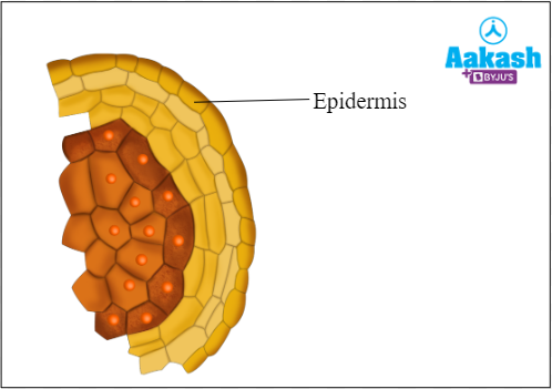
Fig: Epidermis shown in a single microsporangium
Endothecium
The second layer inner to the epidermis is the endothecium. It protects the pollen that is present in the sac and aids in the splitting of anther to release pollen. The cells of the endothecium are large-sized and often develop thickenings of cellulose on the inner and radial walls. The mature cells become dead. This layer is also known as the fibrous layer due to the presence of fibrous thickenings.
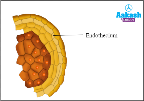
Fig: Endothecium shown in a single microsporangium
The hypodermal as well as epidermal cells present in the region of the shallow groove between the microsporangia of an anther lobe remain thin-walled to function as the stomium or line of dehiscence.

Fig: Stomium
Middle layer
It is the third layer of microsporangium which is composed of 3-4 layers of flattened cells. It lies inner to the endothecium. The cells of the middle layer are short-lived but they may contain starch grains and may also develop some fibrous thickenings. These layers are also protective in nature and help in dehiscence.

Fig: Middle layers shown in a single microsporangium
Tapetum
The innermost layer of the wall of the microsporangium is the tapetum. The tapetum encloses the sporogenous tissue. It provides nourishment to the developing pollen grains. The cells of the tapetum have dense cytoplasm and are multinucleated and polyploid. The tapetum eventually degenerates.
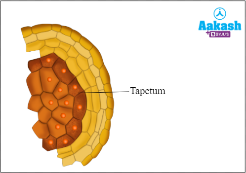
Fig: Tapetum shown in a single microsporangium
The tapetum can be of two types -
- Amoeboid (invasive) - The tapetal cells grow and fuse to form a periplasmodium that passes in between the spore mother cells for providing them with nourishment and other materials. This type of tapetum is seen in lilies, Alisma, Typha, etc.
- Secretory or glandular - Tapetal cells remain in their position, parietal to the sporogenous tissue. They secret nourishment that passes into the sporogenous cells, e.g, Symphoricarpos.
Functions of tapetum
The tapetum not only helps in providing nourishment to the growing sporogenous cells, microspore mother cells and young microspores, but it also secretes hormones that are stored in the pollen grains for their early growth. It provides the enzyme callase that helps to dissolve the callose that binds the microspores. It secretes the Ubisch granules that produce sporopollenin needed to form the outermost exine covering of the pollen grain. It provides the pollenkitt covering around the insect-pollinated pollen grains. It also provides the compatibility-incompatibility proteins to the pollen grains.
Sporogenous tissue
The sporogenous tissue, which is enclosed by the tapetum, contains diploid cells that undergo continuous mitosis to form the diploid microspore mother cells (MMC) or pollen mother cells (PMC). The microspore mother cells are initially connected by plasmodesmata but formation of callose layers on the inner side of the cell wall breaks the plasmodesmata and the microspore mother cells separate out. These microspore mother cells undergo meiosis to form the microspores.
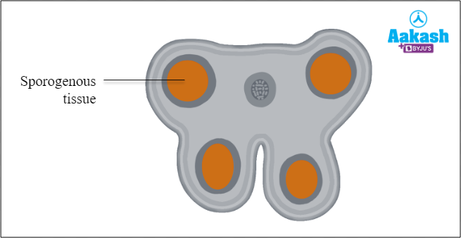
Fig: Sporogenous tissue
Steps involved in microsporogenesis
The process of formation of microspores from sporogenous tissue is known as microsporogenesis. It occurs in the microsporangia of sporogenous tissue. This process involves the following steps:
- Formation of pollen mother cell
- Meiosis I
- Meiosis II
Pollen mother cell
There are several cells present in the sporogenous tissue. Every cell has the potential to develop into microspores or pollen grains. The potential sporogenous cells are known as pollen mother cells or microspore mother cells, or meiocytes. Each pollen mother cell is diploid (2n).

Fig: Pollen mother cell
Meiosis I
The diploid pollen mother cell undergoes first reductional division and this will form two haploid cells.
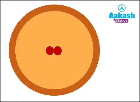
Fig: Meiosis I
Meiosis II
Further, these cells undergo second reduction division and produce two more haploid cells. The total number of haploid cells is four and they are arranged in a cluster. This cluster is known as microspore tetrad. Similarly, all other microspore mother cells present in the sporogenous tissue undergo meiosis I and meiosis II to form tetrads. Therefore, several thousand microspores or pollen grains are produced in each microsporangium. The microspore tetrads can be tetrahedral, isobilateral, decussate, T-shaped or linear.
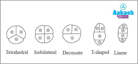
Fig: Microspore tetrad
As the anthers mature and dehydrate, the wall of the microspore mother cell degenerates and the microspores separate and develop into pollen grains. However, in some cases they do not separate and remain united in tetrads, known as compound pollen grains, e.g., Typha, Juncus,
Etc.
In orchids and milkweeds (Calotropis, Asclepias) all the pollen grains of an anther lobe remain united in a sac called pollinium.
Microgametogenesis
The haploid microspores further grow and mature to form haploid pollen which is the male gametophyte. The process of formation of male gametes from the male gametophyte is known as microgametogenesis and it involves several mitotic divisions.
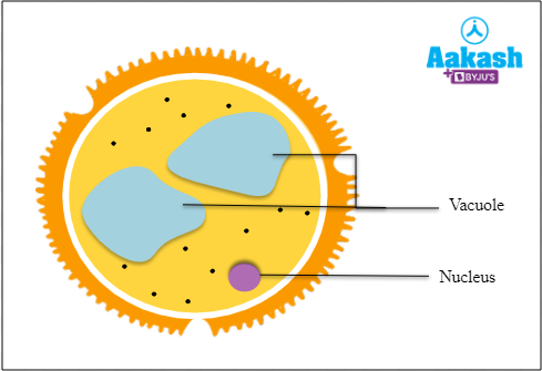
Fig: Structure of microspore
In the newly differentiated pollen grain, the nucleus lies in the centre of a dense cytoplasm which is surrounded by a plasma membrane lying in close contact with the cell wall. The process begins with the expansion of the microspore and a large vacuole is produced. After that, the microspore nucleus moves from the centre to one side of the cell, against the microspore wall. At this location, the first mitotic division of the protoplast takes place. Due to the peripheral position of the nucleus, the mitotic division is asymmetric and gives rise to two unequal cells, a large vegetative cell and a small generative cell, both with haploid nuclei. The pollen at this stage is said to be at a 2-celled stage.

Fig: 2-celled stage
Vegetative cell
The vegetative cell is a large-sized cell which contains small vacuoles and other cell organelles. It has abundant food reserves such as stored scratch, proteins and lipids, to provide nourishment to the developing cell. It also contains unsaturated fatty acids. It has an irregularly shaped nucleus.
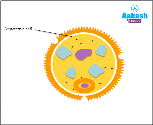
Fig: Vegetative cell
Generative cell
The generative cell is smaller in size as compared to the vegetative cell. It has a spherical or spindle-shaped outline and consists of a thin layer of cytoplasm around a compact nucleus. A layer of callose develops to separate the generative cell from the pollen grain wall. Later, as the callose covering dissolves, the generative cell floats in the cytoplasm of a vegetative cell. In 60% of angiosperms, pollen grains are released at this two-celled stage and further division of the generative cell occurs once the pollen lands on the stigma of a flower and starts germinating.
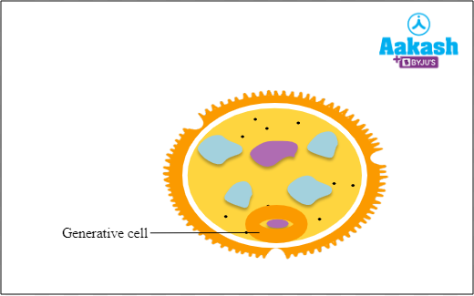
Fig: Generative cell
However, in around 40% of angiosperms, the engulfed generative cell undergoes another mitotic division and forms two pollen sperm cells or male gamete cells that are packed in the cytoplasm. This is now known as the three-celled stage. There are some species of plants that release pollen grains at this stage because pollen grains have completely matured.

Fig: Three-celled stage
Pollen structure
The final mature pollen grain or male gametophyte has a three nuclei stage, two male gametes, and one vegetative nucleus also known as a tube nucleus. The vegetative nucleus later helps in the formation of pollen tube through the germ pore. The pollen is generally spherical in shape and about 25-50 micrometres in diameter. The pollen is covered by a 2 layered wall known as the sporoderm. The two layers of a sporoderm are:
- Intine
- Exine
Exine
The outermost and thick layer of the pollen grain is exine. It is a tough layer because it is composed of sporopollenin. Sporopollenin is related to cutin and suberin and is derived by the oxidative polymerisation of carotenoids. It is one of the most resistant organic compounds that is not affected by high temperature, strong acid, strong alkali, or any known enzyme.
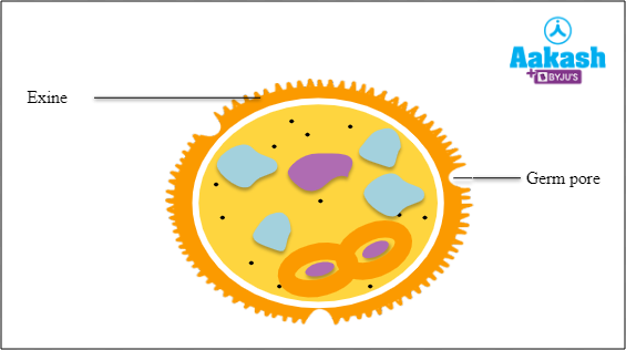
Fig: Exine
The surface of the exine may be smooth, pitted, spiny, warty, etc. The sculptured form of exine is specific for each type of pollen grain. At certain points the exine and hence sporopollenin is absent. These regions are known as germ pores, if rounded or oval areas, or germinal furrows, in case of elongated areas.
The exine of anemophilous or wind-pollinated plants is smooth whereas that of entomophilous or insect-pollinated ones is spiny and covered with a yellowish sticky fatty substance known as pollenkitt. Pollenkitt helps the pollen to stick to the body of the insects. The exine also contains proteins for compatibility-incompatibility reactions and certain enzymes.
The exine is further differentiated into two layers - outer ektexine and inner endexine.
Ektexine
It is the outer sculptured part of the exine and is differentiated into -
- Foot layer - innermost continuous layer of ektexine over which the sculpted part is attached.
- Baculate layer - middle layer of ektexine which is made up of rod-like elements called bacula.
- Tectum - outermost layer which forms a roof above the baculate layer. It can either be smooth or be variously sculptured. Both baculate layer and tectum produce designs all over the pollen surface. This helps in identification of the class, family, genus and species of the plant to which the pollen belongs.
Endexine
It is the inner layer of exine which separates the foot layer of ektexine from the outer, cellulose-rich layer of the intine lying below.
Intine
The inner and thin layer of a pollen grain is known as intine. It is a continuous layer and is composed of cellulose and pectin.
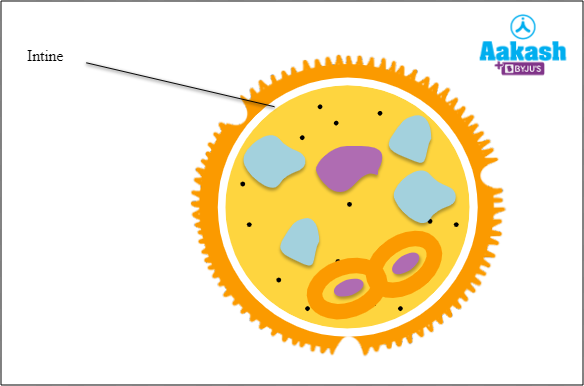
Fig: Intine
Dehiscence of anther to release pollen
Mature anther dries up and the sterile tissue between the two pollen sacs of each anther lobe disintegrates to form a single cavity having a large number of pollen. Thus each mature anther consists of one pollen filled cavity in each of its two anther lobes. Each cavity is now covered only with epidermis and endothecium. The region of the outer thin walls of the dead endothecial cells contract and the cells become concave in shape. The endothecium shortens and the thin-walled cells of the stomium rupture. This produces two longitudinal slits, one on each anther lobe. Thus the anthers are exposed for pollination.
Pollen viability
Pollen viability refers to the pollen's capacity to reach maturity, fertilise, and, following fertilisation, to mature into seeds and fruits. Pollen viability depends on the genetic potential of the plant and the ambient temperature and humidity. Pollen of some cereals can remain viable only for 30 minutes. Consequently, self pollination occurs in such plants. Pollen of flowers belonging to the family Rosaceae, Fabaceae and Solanaceae can remain viable for months. Pollen grains can also be cryopreserved in liquid nitrogen at -196oC and be used for breeding purposes.
Pollen products
Pollen is a rich source of vitamins and therefore, it is used in food supplements and pollen creams.
Pollen food supplements
Pollen grain contains a variety of nutrients, such as proteins, carbohydrates and unsaturated fats. Due to these nutrients, pollen is used in food supplements in the form of tablets and syrups. The food supplements that contain pollen enhance the vital functions of the body. Therefore, they are used to increase the performance of race horses and athletes.
Pollen creams
Pollen grains contain unsaturated fats that protect them from UV rays. Due to this property, pollen is used in creams, emulsions and cosmetics to provide protection against the UV rays.
Significance of microsporogenesis
Microsporogenesis is significant because it helps to maintain the ploidy of the gamete. The microspore mother cells or meiocytes are diploid cells that undergo meiosis to produce four haploid microspores which develop into the male gametophyte containing two haploid male gametes. The haploid male gamete fertilises the haploid egg cell of the female gametophyte and forms a diploid zygote which will grow into a new plant. Thus, the diploid chromosome number is restored in the offspring.
Meiosis during microsporogenesis also allows random segregation and crossing over of chromosomes which helps in bringing about variations in the genetic composition of the male gametes compared to that of the parent plant.
Microsporogenesis vs Megasporogenesis
|
Microsporogenesis |
Megasporogenesis |
|
The process through which micropores are produced from the microspore mother cell through reductional division is known as microsporogenesis. |
The process through which megaspores are formed from the megaspore mother cell is known as megasporogenesis. |
|
The process of microsporogenesis occurs in the pollen sac of an anther. |
The process of megasporogenesis occurs in the ovule of an ovary. |
Practice Problems
- Identify the structure that directly develops into male gametophyte in angiosperms.
- Microspore
- Microsporangia
- Sporogenous tissue
- Endothecium
Solution: In angiosperms, microspores are haploid plant spores that transform into male gametophytes (pollen grains). Hence, the correct option is a.
2. Identify the odd one out with respect to the ploidy of a cell.
- Pollen mother cells
- Tapetum
- Endothecium
- None of the above
Solution: The innermost layer of the anther wall is tapetum. It is rich in food reserve and therefore, provides nourishment to the developing pollen grains. The cells of the tapetum are called tapetal cells and contain dense cytoplasm with more than one nucleus. Therefore, it is polyploid. Pollen mother cells and endothecium are diploid in nature. Hence, the correct option is b.
3. Microsporangia or ______i________ in anther have microspores that develop into ______ii_______.
- i-pollen grains, ii-pollen sacs
- i-pollen sacs, ii-male gametophytes
- i-embryo sac, ii-pollen grains
- i-male gametophytes, ii-embryo sacs
Solution: Microsporangia is described as a sac-like structure that possesses microspores. It is also known as the pollen sac. The microspores in the pollen sac develop into pollen grains or male gametophytes. Hence, the correct option is b.
4. Identify the structure that is present at the centre of the young anther.
- Tapetum
- Endothecium
- Sporogenous tissue
- None of the above
Solution: A homogenous mass of meristematic cells is present in the young anther and this mass is known as sporogenous tissue. It is located in the centre of the young anther and is surrounded by the anther wall. These cells produce microspore mother cells that develop into male gametophytes or pollen grains. Hence, the correct option is c.
FAQs
- What gives yellow colour to pollen?
Answer: Most of the plant species have yellow pollen due to the presence of flavonoids, which are known to have UV-B protecting effects.
- Do all plants produce yellow pollen?
Answer: Most plants produce yellow pollen but not all. Pollen can also come in other colours such as red, purple, brown, etc. Since most insects cannot detect the red colour, insect pollinated flowers produce yellow or blue pollen to attract the insects. On the other hand, birds are attracted by red colour and ornithophilous or bird pollinated flowers produce red pollen.
- What are the common symptoms of pollen allergy?
Answer: The common symptoms of pollen allergy are running nose, red eyes, sneezing, cough, and itchy throat.
- Why is carrot grass harmful for humans?
Answer: Carrot grass or Parthenium is an exotic plant species which came to India along with imported wheat species and is presently one of the major causes of weed pollen allergies. It releases sesquiterpene lactones in large amounts which causes skin allergies.
Youtube link: https://www.youtube.com/watch?v=DVbLRuXGlko