Full Form of VSD in Medical: In medical terms, VSD stands for Ventricular Septal Defect. It is a congenital heart condition, which means it is present at birth. In this defect, there is a hole in the wall (septum) that separates the two lower chambers of the heart, known as the left and right ventricles.
This opening allows oxygen-rich blood from the left side to mix with oxygen-poor blood on the right side. As a result, the heart has to work harder to pump enough oxygen to the body. VSD is one of the most common types of congenital heart diseases found in newborns.
Understanding the Heart and VSD
The heart is a muscular organ that pumps blood throughout the body. It has four chambers—two upper chambers (right and left atria) and two lower chambers (right and left ventricles). These chambers are separated by walls called septa. The ventricular septum separates the left and right ventricles and plays a key role in preventing the mixing of oxygen-rich and oxygen-poor blood.
The full form of VSD is Ventricular Septal Defect. When a VSD occurs, there is a hole in the ventricular septum. This opening allows blood to flow abnormally from the left ventricle to the right ventricle. As a result, too much blood flows to the lungs, increasing the pressure in lung arteries and making the heart work harder. Over time, if the defect is large and untreated, it can cause serious heart and lung problems.
Types of Ventricular Septal Defect
VSDs are classified based on where the hole is located in the ventricular septum and how large the hole is. The main types include
-
Perimembranous VSD
This is the most common type. The hole is found in the upper section of the septum, near the heart valves. These VSDs may need surgery if they are large or cause symptoms. -
Muscular VSD
This type is located in the lower muscular part of the septum. These VSDs often close on their own as the child grows older. They are usually less serious and might not require surgery. -
Inlet VSD
Inlet VSDs are located near the tricuspid and mitral valves, where blood flows into the heart. These are sometimes associated with other heart conditions. -
Outlet VSD (or Supracristal VSD)
This is a rare type where the hole is located near the blood vessels (aorta and pulmonary artery). These VSDs are more common in certain Asian populations and often need surgical repair.
Causes of VSD
The exact cause of VSD is not always known, but several factors may lead to its development:
-
Genetic causes: Sometimes, VSD can be inherited or associated with genetic disorders like Down syndrome or other congenital conditions.
-
Environmental exposure: If a pregnant woman is exposed to certain infections (like rubella) or harmful substances such as alcohol or drugs, it may affect the development of the baby’s heart.
-
Improper heart development: VSD happens when the baby’s heart does not form properly in the womb during the first 8 weeks of pregnancy.
It is important to note that in many cases, there is no known cause for the defect.
Symptoms of VSD
The signs and symptoms of VSD depend on the size of the hole and how much blood passes through it.
-
Small VSD: May cause no symptoms at all. Many children with small VSDs appear completely healthy.
-
Medium to large VSDs: May cause the following symptoms, especially in infants:
-
Shortness of breath or trouble breathing, especially during feeding
-
Sweating while eating or crying
-
Frequent chest infections or lung infections
-
Fatigue or weakness
-
Poor weight gain or slow growth
-
Fast heartbeat or irregular heart sounds (murmurs)
-
If these symptoms are noticed, a doctor should be consulted immediately for a proper heart evaluation.
Diagnosis of VSD
VSD is usually detected in infants or young children. In some cases, it may be discovered later during a health check-up. Common ways doctors diagnose VSD include
-
Physical Examination: A doctor may hear a heart murmur, which is an unusual sound caused by the blood flowing through the hole in the heart.
-
Echocardiogram (Echo): This test uses sound waves to produce a moving image of the heart. It clearly shows the location and size of the VSD and how blood is flowing.
-
Electrocardiogram (ECG or EKG): This test checks the electrical activity of the heart and helps detect signs of heart enlargement.
-
Chest X-ray: It shows the size and shape of the heart and helps identify any fluid buildup in the lungs.
-
Cardiac MRI or cardiac catheterization: In some rare cases, these advanced tests may be used to get a clearer picture of the defect.
Treatment Options for VSD
Treatment depends on several factors, including the size of the VSD, the age of the patient, and the symptoms. The main treatment options are
-
Observation and Monitoring
If the VSD is small and symptomless, the doctor may simply watch and wait. Regular follow-ups and tests are done to ensure the hole is closing on its own or not causing any problems. -
Medications
For moderate symptoms, medicines can help control them. Commonly used medications include:-
Diuretics (to remove extra fluid and reduce heart workload)
-
ACE inhibitors (to lower blood pressure and make heart pumping easier)
-
Digoxin (to strengthen heart contractions)
-
-
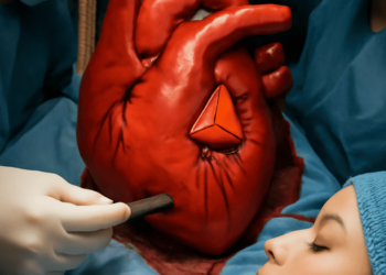
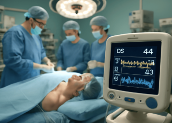
Surgery
If the VSD is large or causes severe symptoms, open-heart surgery may be recommended. The surgeon closes the hole using a patch or stitches. This is often done between 3 and 12 months of age, depending on the child’s condition. -
Catheter Procedure
In some cases, especially for older children or adults, a thin tube called a catheter is inserted through a vein to the heart. A device is placed to close the hole without the need for open surgery.
Complications of Untreated VSD
If a large or symptomatic VSD is left untreated, it can lead to serious long-term health problems, such as
-
Heart failure: The heart cannot pump enough blood to meet the body’s needs.
-
Pulmonary hypertension: Increased blood flow to the lungs can damage lung arteries.
-
Endocarditis: Infection in the heart’s inner lining, which can be life-threatening.
-
Arrhythmia: Abnormal heart rhythms due to the stress on heart muscles.
-
Delayed growth and development: Children may grow slowly or have poor physical development.
That’s why early diagnosis and timely treatment are very important.
Living with VSD
Most children born with a small VSD live completely normal lives without any complications. If the defect closes on its own or is treated successfully, the child may not need any further care apart from routine checkups.
For children who have undergone surgery, recovery is usually very good. They may be asked to avoid certain activities for a short period and may require regular heart check-ups into adulthood. Good oral hygiene is also important to prevent infections like endocarditis.
In general, with proper treatment and follow-up, people with VSD can lead healthy, active lives.
Conclusion
The full form of VSD in medical terms is Ventricular Septal Defect. It is a common heart defect that happens due to a hole in the wall separating the heart’s lower chambers. The condition may be minor and harmless or serious and require surgery. However, modern medical techniques make it possible to treat VSD effectively, and most patients recover well and live normal lives.