-
Call Now
1800-102-2727


Meristematic Tissue, Practice Problems and FAQs
Gardening is a common hobby amongst many people. Maintaining a good garden is difficult and requires a lot of effort. You might have seen some bushy plants maintained very well in most of the gardens. People cut bushes in different designs for aesthetic beauty. What they commonly do is cut the apical portions of the plant to inhibit the growth. But why do the plants stop growing when we cut them from the apex?
You know that growth occurs because of the division of cells. In plants the active cell division happens in some regions and the tissues of those regions are responsible for the growth in plants. Let’s understand this through an experiment.
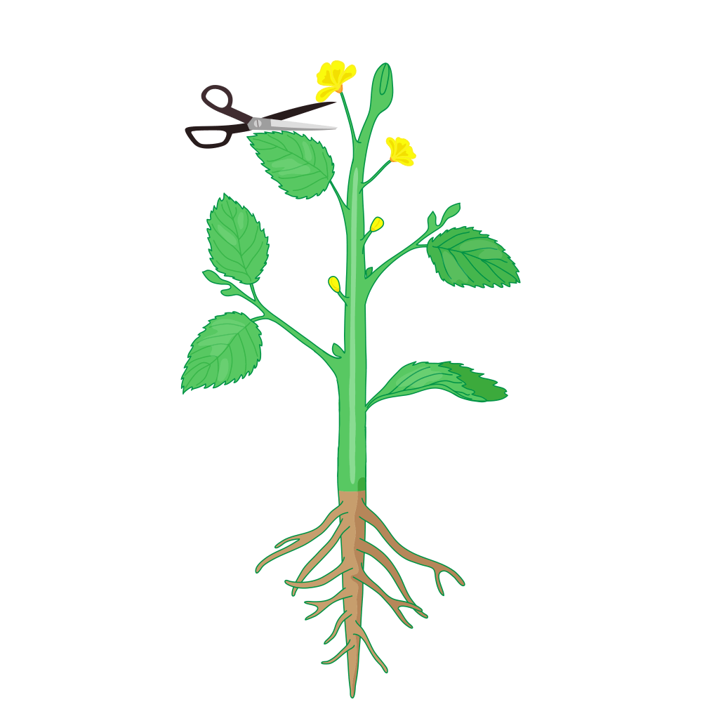
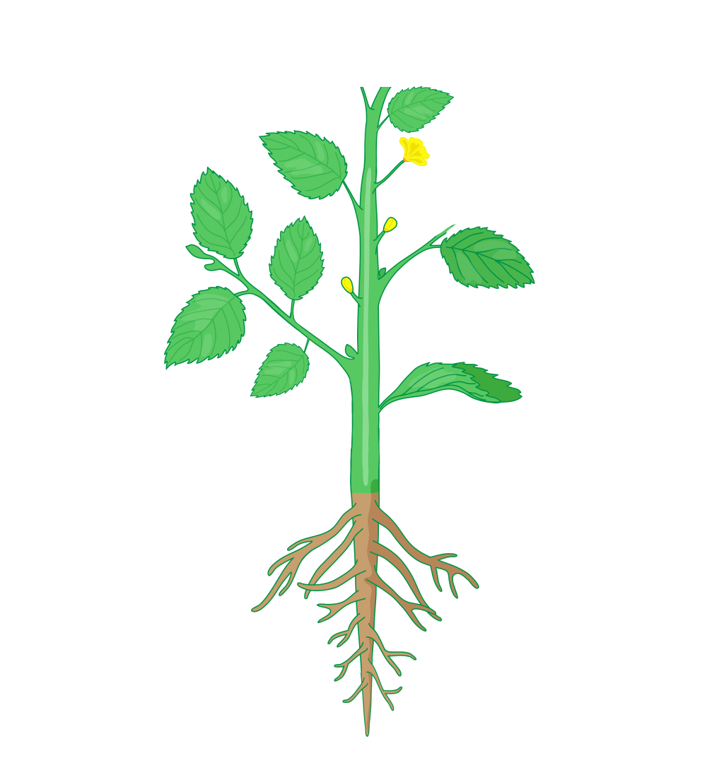
In the first scenario, let us cut off the tip of the main stem of a plant. Here the branches and roots will grow, but the main stem will not grow.
GIF: Cutting the main GIF: No growth in the main stem
stem of the plant
In the second scenario, let us cut off the tips of some of the side branches of the plant. Here the main stem and main root will grow, but the branches will not grow.
GIF: Cutting the side branches GIF: No growth in the


of the plant side branches
In the third scenario, let us cut off the tip of the root of the plant B. Let's see what happens. Here the branches will grow, the main stem will grow but the main root will not grow.
GIF: Cutting the tip of root GIF: No growth in the main root


Now let us compile all the data. In each of the above scenarios we see that when the tips of the plant parts are cut off, the growth in those regions is inhibited. This is because the tips of plant parts contain a tissue called meristematic tissue which has actively dividing cells that contribute to growth. Meristematic tissue can be classified into different types based on different criterias. In this article we are going to discuss more about the meristematic tissues in plants.
Table of contents:
- Tissues
- Meristematic tissues
- Classification of meristematic tissues
- Structural organisation of apical meristems
- Theories of the structural organisation of apical meristems
- Functions of meristematic tissues
- Practice Problems
- FAQs
Tissues
The group of similar or dissimilar cells that work together to perform a specific function and also have the same origin are called tissues.
Types of plant tissues
On the basis of capacity to divide, there are two types of plant tissues. They are as follows:
- Meristematic tissues
- Permanent tissues
Fig: Classification of plant tissues

Meristematic tissues
Meristems are the specialised regions in the plant where the active cell division takes place and the growth is restricted to this region. The tissues present in the meristem are called meristematic tissues. These tissues are a group of immature and similar cells. They are also undifferentiated cells which remain in the state of continuous division.
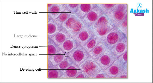
Characteristics of meristematic tissues
- Cells are immature and undifferentiated and are in a state of active division and continuous growth.
- Cells are thin-walled, isodiametric and compactly arranged, usually without intercellular space.
- Protoplasm is dense with one or more prominent nuclei.
- Cells do not store reserve food materials.
- Vacuoles are very small or absent.
- Plastids exist as protoplastids.
- Cell wall is thin and primary and is formed of only cellulose.
Fig: Meristematic tissue
Classification of meristematic tissues
Meristems are classified on the basis of their position in the plant body, origin and development, the plane of cell division and function.
Classification of meristems based on origin and development
Based on the origin and method of development, meristems are classified into three types. They are as follows:
- Promeristem
- Primary meristem
- Secondary meristem

Pro meristem
Those meristems which are embryonic in origin are called pro meristems or primordial meristems or embryonic meristems. It occurs in the regions where the development of an organ or a plant part is initiated. It is present in the germinating embryos or young seedlings. In embryos it is located at the tips of plumule and radicle and in a growing plant it is seen at the tips of roots and shoots.
Promeristem consists of initials and their immediate derivatives. Its cells are isodiametric and thin walled, with prominent nucleus, active cytoplasm and early stages of pit formation. Intercellular spaces are usually very inconspicuous.
Fig: Promeristem

Primary meristem
The meristem originates from the promeristem during the embryonic stage and builds up the primary part of the plant body is called primary meristem. It is seen at the apices of roots, stems and leaf primordia. It is concerned with the formation of the primary permanent tissues of various organs.
Functionally, primary meristem builds up the structurally and functionally complete initial plant body to which secondary meristem subsequently adds supplementary tissues. These supplementary tissues functionally replace the primary ones or protect and repair the regions. Examples of primary meristem are as follows:
- Apical meristem
- Intercalary meristem
- Intrafascicular cambium
Fig: Primary meristem

Secondary meristem
The meristem develops from non-meristematic primary permanent tissues at a later stage of development by regaining the power for active cell division is called secondary meristem. Secondary meristems are formed through dedifferentiation and help in the increase in the girth of a plant. So, secondary meristems always arise from permanent plant organs whenever and wherever they are needed. For example vascular cambium originates from interfascicular regions in the dicots during secondary growth and the cork cambium arises from the cortex during periderm formation. Sometimes, some permanent tissues acquire the power for cell division and become meristematic and then give rise to secondary meristems. The examples of secondary meristem are as follows:
- Interfascicular cambium
- Vascular cambium
- Cork cambium
Fig: Secondary growth

Classification of meristems based on the position
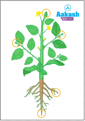
Based on the position in the plant body, meristems are classified into three types. They are as follows:
- Apical meristem
- Intercalary meristem
- Lateral meristem

Apical meristems
The primary meristem located at the growing apices of axillary buds, stems and roots are called apical meristems. They are formed from the pro meristems. It is responsible for the linear growth or elongation of plant organs, such as shoot tip and root tip. By the activity of the apical meristems of roots and shoots all the primary tissues of the plant body are formed.
The actively dividing cells of the apical meristem are called apical initials or apical meristematic cells. They are located at the extreme tip of the roots and shoots. In pteridophytes, apical initials are solitary cells. But in higher vascular plants they occur in groups which may be terminal or subterminal in position.
Fig: Apical meristems
Types of apical meristems
There are two types of apical meristems and they are as follows:
- Shoot apical meristems
- Root apical meristems
Shoot apical meristems
The apical meristem at the shoot apex is called shoot apical meristem. They are terminal in position.

GIF: Shoot apical meristems
Primary tissues are produced by the shoot apical meristem. During the formation of leaf and elongation of stem, some tissues are left behind from the shoot. From these tissues axillary buds are formed. A branch or flower can form from the axillary buds.
Fig: Axillary buds

Root apical meristems
The apical meristems that are seen on the root apex, which are subterminal in position are called root apical meristems. The elongation of the roots are done by the root apical meristem.

Intercalary meristems
During the early stages of the development, internodes are wholly or partially meristematic. Intercalary meristems are the internodal meristems that present between the mature tissues. They are actually the portions of the apical meristems that are left behind at the nodal regions during tissue differentiation.
Fig: Intercalary meristems
They are responsible for the elongation of the internodal regions and are seen mostly at the stem nodes of many monocots such as sugarcane, paddy, wheat and many grasses.

GIF: Elongation of internodes
Intercalary meristems are commonly found in between masses of permanent tissues either at the leaf base or at the base of the internodes. They are short living because they become permanent tissues after a period of division, growth and differentiation.

Lateral meristems
The meristem occupying the lateral side of root and stem are called lateral meristems. It consists of initials which divide mainly in one plane or periclinal. This causes an increase in the girth during secondary growth. Usually it adds to the bulk of the existing tissues, or forms new tissues. Lateral meristem is absent in monocot stems. They are called secondary meristems, since they are formed after primary meristems.
Fig: Lateral meristems

The examples of lateral meristems are as follows:
- Vascular cambium (interfascicular and interfascicular cambium)
- Cork cambium
- Wound cambium
Lateral meristems are also called cylindrical meristems, because of their shape.
Fig: Cylindrical meristems

Classification of meristems based on the plane of division
On the basis of the plane of the cell division, meristems are classified into three and they are as follows:
- Rib meristem
- Plate meristem
- Mass meristem
Rib meristem
If the cells are dividing anticlinal in the meristematic tissue, then it is known as rib meristem. It plays an important role in the development of cambium and cork cambium.
Plate meristem
The meristematic tissue by which the cells dividing both anticlinal and periclinal is called plate meristem. This will give rise to a plate-like increase in cell mass. It plays an important role in the development of leaves.
Mass meristem
The meristematic tissue whose cells divide in all planes are called mass meristem. This results in massive tissue development. It plays an important role in the development of endosperm, sporangia, cortex and pith.
Classification of meristematic tissues on the basis of function
Based on the functions of the meristematic tissues, they can be classified into three by Haberland and they are as follows:
- Protoderm
- Procambium
- Ground meristem
Protoderm
The outer primary meristem of the plant is called protoderm. The function of protoderm is to protect the plants against mechanical shocks.

Procambium
The deepest tissue which produces xylem and phloem is called procambium. The function of procambium is to help in the movement of water and nutrients in the plant.

Ground meristem
The tissue that can generate all cells of a plant except the epidermis and vascular tissues is called ground meristem. It can undergo rapid cell division.
Structural organisation of apical meristems
Growth takes place by the activity of a group of initial cells in vascular plants. These initial cells originate from embryonic root and shoot and are present at the tips of the shoot and root. This forms the apical meristems. The structural organisation of root apex and shoot apex are different.
Structural organisation of root apex
A typical dicot root apex has four distinct regions. They are as follows:
- Calyptra or root cap
- Calyptrogen
- Quiescent centre
- Meristematic region
The apical meristem together with the root cap is called root apex. It is embryonic in origin and is responsible for the formation of all the tissues of the primary root.
Calyptra or root cap
The outermost protective layer of the root apex is called calyptra or root cap. It is formed of parenchyma cells that are short-living. It helps the root to penetrate deep into the soil and rock, through secreting a lubricant substance. Starch grains are present in the parenchyma cells.
Calyptrogen
The region within the root apical meristem, located just inner to the root cap is called calyptrogen. It is a group of actively dividing cells, which divide and produce calyptra.
Quiescent centre
A central cap-like or hemispherical region of inactive cells within the root apical meristem is called quiescent centre. It is located in between the calyptrogen and the active meristematic region. It consists of a group of slowly dividing cells, surrounded by actively dividing cells. It is normally inactive.
Meristematic region
This is the region where the cells are divided repeatedly and the primary root tissues emerge.

Structural organisation of shoot apex
The location of the shoot apex is at the tip of the stem and it branches as terminal buds. It is in an inactive state in the axils of the leaves as lateral buds. It is conical or dome shaped and is always covered by young leaves, arising from its sides. But shoot apices can be highly variable in size and shape. The apical meristem becomes broader before the initiation of each leaf. But after giving out the leaves, it again becomes narrow.
Theories of the structural organisation of apical meristems
Several theories have been proposed to explain the structure and organisation of root and shoot apex. The most widely accepted ones are apical cell theory, the Korper-Kappe theory, tunica-corpus theory and histogen theory. In this the Korper-Kappe theory is exclusive for root and tunica-corpus theory is exclusive for shoot.
Apical cell theory
This theory was proposed by Nageli in 1858. It states that the apical meristem of root and shoot consists of a single apical cell and the growth and development of the entire plant body result from the activity of this cell. The apical cell theory is applicable to higher algae, bryophytes and many pteridophytes. But it is not applicable in the case of gymnosperms and angiosperms. Recent studies using colchicine have confirmed that the shoot apex of gymnosperms and angiosperms consists of a group of cells which constitute the meristem.
Tunica-corpus theory
This theory was proposed by Schmidt in 1924 to describe the shoot apex. According to this theory, shoot apex consists of two distinct zones, namely outer tunica and inner corpus. Tunica surrounds and envelopes the central corpus. The cells of tunica are smaller than those of corpus. Tunica cells divide anticlinaly so that the number of layers does not increase. On the other hand, corpus cells divide anticlinaly and periclinaly.
The outer tunica gives rise to the epidermis, while its inner layers give rise to the outer part of the cortex. The cells of the corpus give rise to the inner part of the cortex and also the procambium and pith. The procambium, in turn, gives rise to the primary phloem and xylem. Thus shoot apex gives rise to all the primary tissues of the shoot system. During the flowering stage, it gives rise to the floral leaves like sepals, petals, carpels and stamens also. The shoot apex which produces the floral leaves, instead of leaf primordia, is often called the floral apex.

according to tunica-corpus concept
Korper-Kappe theory
This theory was advanced by Schuepp in 1917. It holds that each cell at the root apex divides in two planes. The first division is transverse and then one of the daughter cells divides longitudinally. The sequence of divisions is called T-division, because cell walls form a T-shaped configuration.
In some zones of the root, especially in the centre T-shape is erect or straight T, whereas in other regions it is inverted T. In the erect condition, the bar of the T faces the root apex, whereas in the inverted condition it faces away from the root apex. The zone with inverted T type division is termed korper or cap and the one with straight T-type division is called kappe or body. This theory is basically similar to the tunica-corpus concept of shoot apex. As a whole, it fails to explain the differences in behaviour in different species.

Histogen theory
The histogen theory was proposed by Hanstein in 1870. This theory holds that apical root and shoot meristem consists of different meristematic zones or layers called histogens. There are usually three or four groups of initials in angiosperms. In dicots, there are three layers, and they are dermatogen, periblem and plerome. Each layer performs definite functions. Dermatogen forms the epidermis in all organs of the plant. Periblem gives rise to cortex and endodermis. Plerome forms the pith and primary vascular tissues.
Functions of meristematic tissue
The major functions of meristematic tissues are as follows:
- Active division of plant tissues happens at the meristematic regions.
- New organs are produced through the division of meristematic tissues and it also supports the development of new organs in the plants.
- The formation of secondary tissues like wood and cork is by meristems and these tissues support the secondary growth of a plant.
- If root development is interrupted or root tip is wounded, then the meristematic tissue recovers the growth.
Practice Problems
1. Which of the following statements are wrong about the meristematic tissues of a plant?
- The immature and undifferentiated cells present in the meristematic regions are called meristematic tissues.
- Meristematic cells are thin-walled, isodiametric and compactly arranged.
- Meristematic tissues have intercellular spaces.
- Meristematic cells do not store reserve food materials.
Solution: The group of similar or dissimilar cells that work together to perform a specific function and also have the same origin are called tissues. On the basis of capacity to divide, there are two types of tissues. They are meristematic tissues and permanent tissues. Meristems are the specialised regions in the plant where the active cell division takes place and the growth is restricted to this region. The tissues present in the meristem are called meristematic tissues. These tissues are a group of immature and similar cells. They are also undifferentiated cells which remain in the state of continuous division. Meristematic tissues have cells that are thin-walled, isodiametric and compactly arranged, usually without intercellular space. Protoplasm is dense with one or more prominent nuclei in meristematic cells and they do not store reserve food materials. Vacuoles are very small or absent in the cells. Plastids of meristematic tissues exist as protoplastids. Cell wall is thin and primary and is formed of only cellulose. Hence the correct option is c.
2. Which of the following rows are correctly paired?
|
A |
Primary meristem |
Apical meristem, Intercalary meristem, Intrafascicular cambium |
|
B |
Secondary meristem |
Interfascicular cambium, Vascular cambium, Cork cambium |
|
C |
Lateral meristem |
Vascular cambium, Cork cambium, Wound cambium |
- A only
- B and C
- A and B
- A, B and C
Solution: The meristem originates from the promeristem during the embryonic stage and builds up the primary part of the plant body is called primary meristem. It is seen at the apices of roots, stems and leaf primordia. It is concerned with the formation of the primary permanent tissues of various organs. The examples of primary meristem are apical meristem, intercalary meristem and intrafascicular cambium. The meristem develops from non-meristematic primary permanent tissues at a later stage of development by regaining the power for active cell division is called secondary meristem. Secondary meristems are formed through dedifferentiation and help in the increase in the girth of a plant. The examples of secondary meristem are interfascicular cambium, vascular cambium and cork cambium. The meristem occupying the lateral side of root and stem are called lateral meristems. It consists of initials which divide mainly in one plane or periclinal. This causes an increase in the girth during secondary growth. The examples of lateral meristems are vascular cambium, cork cambium, wound cambium. Hence the correct option is d.
3. Which of the following meristems initiate the formation of a flower or branch?
- Shoot apical meristem
- Root apical meristem
- Lateral meristem
- Rib meristem
Solution: The primary meristem located at the growing apices of axillary buds, stems and roots are called apical meristems. They are formed from the pro meristems. It is responsible for the linear growth or elongation of plant organs, such as shoot tip and root tip. There are two types of apical meristems and they are shoot apical meristems and root apical meristems. The apical meristem at the shoot apex is called shoot apical meristem. They are terminal in position. Primary tissues are produced by the shoot apical meristem. During the formation of leaf and elongation of stem, some tissues are left behind from the shoot. From these tissues axillary buds are formed. A branch or flower can form from the axillary buds. Hence the correct option is a.
4. Which of the following theories explains the T shaped configuration of cells in the structural organisation of meristems?
- Apical cell theory
- Korper-Kappe theory
- Tunica-corpus theory
- Histogen theory
Solution: Several theories have been proposed to explain the structure and organisation of root and shoot apex. The most widely accepted ones are apical cell theory, the Korper-Kappe theory, tunica-corpus theory and histogen theory. In this the Korper-Kappe theory is exclusive for root and tunica-corpus theory is exclusive for shoot. Korper-Kappe theory was advanced by Schuepp in 1917. It holds that each cell at the root apex divides in two planes. The first division is transverse and then one of the daughter cells divides longitudinally. The sequence of divisions is called T-division, because cell walls form a T-shaped configuration. In some zones of the root, especially in the centre T-shape is erect or straight T, whereas in other regions it is inverted T. In the erect condition, the bar of the T faces the root apex, whereas in the inverted condition it faces away from the root apex. The zone with inverted T type division is termed korper or cap and the one with straight T-type division is called kappe or body. Hence the correct option is c.
FAQs
1. What is apical dominance?
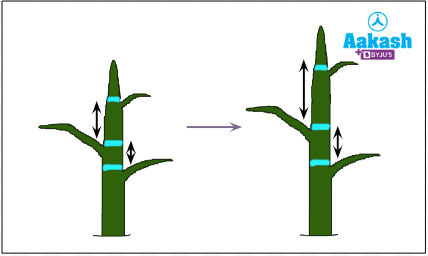
Answer: The inhibition of growth of one meristem by another meristem is called apical dominance. This will result in the formation of only one main trunk in the plants. The tip of the trunk of a tree grows rapidly without being shadowed by branches. Apical dominance is the reason for this, because in trees, the dominant shoot meristem is present in the tip of the main trunk. If this dominant meristem is cut, then there will be more branch tips. This will grow faster than the normal branches and it will look like the extension of the main trunk. This kind of growth will lead to a bushy growth.
2. Why is apical meristem culture important in tissue culture?
Answer: Apical meristem culture is the method of propagating plants in vitro by using the apical meristems with one to three leaf primordia. The major advantage of this technique is that it can create virus free plants, because the meristem is free from viruses. Since meristem has continuously dividing cells, it will have a high metabolic rate. Viruses can not replicate in those cells with high metabolism.
3. What is plastochron?
Answer: If the temperature is constant then new leaves are produced at regular intervals as the tip of shoot grows. The time interval between the growth of new leaves is called plastochron. This is used for the calculation of the age of a plant.
4. Which are the major hormones present in the meristem?
Answer: The two major hormones involved in the function and maintenance of meristem are auxin and cytokinins. Auxins are usually produced in the apical meristem and help in the apical dominance. The major function of cytokinin is the cell proliferation and meristem maintenance in shoot.
YOUTUBE LINK: https://www.youtube.com/watch?v=-rGetleD-DI
https://www.youtube.com/watch?v=ISN_MHEe4Yc
Related Topics
|
The tissues: Meristematic tissues, Classification based on origin, Practice Problems and FAQs |
|
The tissues: Meristematic tissue, Classification based on position, Practice Problems and FAQs |
|
Permanent tissues: Types of permanent tissues, Simple tissues, Practice Problems and FAQs |
|
Permanent tissues: Xylem, Practice Problems and FAQs |